
By EIV Diagnostics · September 16, 2026
Clinicians: Clinicopathologic Correlation Led to 13 Revised Diagnoses
Clinicopathologic correlation improves diagnostic accuracy. See the audit evidence, follow a five-item checklist for biopsies, and apply practical lab...
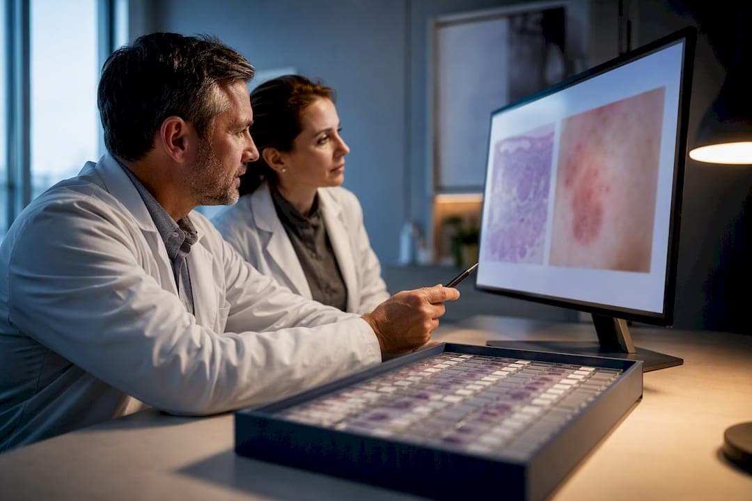
Clinicopathologic correlation (CPC) is the deliberate process of matching a patient’s history, exam findings, and lab or imaging results against what a pathologist sees under the microscope. It sharpens diagnostic confidence and, in a measurable share of cases, changes the diagnosis or the treatment plan entirely. A dermatopathologist reading an ambiguous melanocytic lesion without knowing the patient’s age or lesion history is guessing with one eye closed. A biopsy read alongside a clear clinical note rarely leaves that kind of doubt.
TL;DR:
- Proper specimen handling, including accurate labeling and timely fixation, is critical to preserve tissue architecture and reduce diagnostic errors.
- Including detailed clinical history, provisional diagnosis, and relevant images with every specimen significantly improves diagnostic accuracy and consistency.
- Discrepancies between clinical and pathology findings often stem from missing information or sampling issues, which can be mitigated through re-biopsy or second opinions.
- Routine review and documentation of discordant cases, along with direct communication between clinicians and pathologists, enhance diagnostic reliability over time.
- Digital pathology and secure communication platforms support faster, more accurate clinicopathologic correlation, especially when combined with clinical context and ancillary testing.
Table of Contents
- What Are the Core Steps in Clinicopathologic Correlation?
- What Should You Send With Every Specimen?
- What Causes Discordance Between Clinical and Pathology Findings?
- Does CPC Actually Change Diagnoses? Here’s the Evidence
- How Does a Modern Pathology Lab Support the CPC Workflow?
- How Should You Handle Confidentiality When Sharing CPC Data?
- What Role Do AI and Digital Pathology Play in CPC?
- How Can Departments Improve Communication Around CPC?
- How Do You Audit and Improve CPC Quality Over Time?
- Teaching CPC So It Actually Sticks
- How EIV Diagnostics Supports Your CPC Workflow
- Sources
- FAQ
What Are the Core Steps in Clinicopathologic Correlation?
CPC is not a single moment where a pathologist glances at a chart. It is a workflow with distinct checkpoints, and skipping any one of them weakens the final report.
Specimen handling comes first. Proper fixation, accurate labeling, and, where relevant, gross photography preserve the tissue architecture pathologists depend on. A mislabeled specimen or a delay in fixation can distort morphology enough to throw off an otherwise straightforward read. From there, the pathology side takes over: gross description, tissue processing, microscopic examination, and, when indicated, immunohistochemistry or molecular testing to resolve ambiguous morphology.
None of that happens in a vacuum. Clinical history, prior lab values, imaging results, and clinical photographs get folded into the interpretation at the point the pathologist writes the report, not as an afterthought. This integration is what separates a technically accurate description of tissue from a diagnosis that actually fits the patient in front of you. Documentation practices, formalin fixation timing, and correlating gross findings with the patient’s history are the specific components that make this integration work rather than just happen on paper, according to a teaching-focused review of CPC.
Operationally, this depends on infrastructure most clinicians never think about:
- Requisition forms structured to force a minimum clinical data set, not just demographics.
- Digital systems that let clinicians attach photos or dermoscopic images directly to the order.
- Clear turnaround expectations so urgent cases get flagged rather than queued normally.
Pro Tip: Write your clinical impression as a differential, not a single guess. “Rule out melanoma vs. dysplastic nevus” gives the pathologist something to correlate against; “check this mole” gives them nothing.
What Should You Send With Every Specimen?
Most diagnostic discordance traces back to what never made it onto the requisition form. Here is the minimum a pathologist needs to correlate properly:
- Patient demographics and site. Age, sex, and exact anatomic location, since the same histology reads differently on a sun-exposed cheek than on a shielded trunk.
- A provisional clinical diagnosis or differential. State what you suspect, not just what you removed.
- Duration and evolution. How long has the lesion or lump been present, and has it changed?
- Prior treatment or biopsy history. Steroid creams, prior excisions, and radiation all alter tissue appearance.
- Relevant labs and imaging, summarized. One sentence beats a scanned chart nobody has time to read.
Clinical and dermoscopic images are worth the extra minute it takes to attach them. Label each one with the site and date, and if you are submitting more than one lesion, make sure the image maps unambiguously to the correct specimen container. Studies on pigmented skin tumors found that image-aided review measurably raises diagnostic confidence and occasionally prompts re-evaluation of borderline findings.
Pro Tip: If a case feels genuinely uncertain, say so on the form and ask for an expedited review or a spot at the next CPC conference. Pathologists would rather field that request than issue a diagnosis they later have to walk back.
What Causes Discordance Between Clinical and Pathology Findings?
Most mismatches come from a short list of repeat offenders: a requisition with no real history, a biopsy that missed the diagnostic portion of the lesion, a blurry or mislabeled photo, or a clinician leaning on one test result as if it settles the whole picture.
The fix usually isn’t complicated, just deliberate:
- Re-biopsy or request deeper, targeted tissue blocks when the sample looks non-representative.
- Order immunostains or molecular panels aimed at the specific differential, not a scattershot workup.
- Get a second pathology opinion when the clinical picture and the report genuinely do not line up.
- Document the discordance and its resolution in the chart, so the next clinician isn’t starting from zero.
Closing the loop matters more than any single test. A phone call between clinician and pathologist often resolves in five minutes what a stack of addenda would take a week to sort out.
Does CPC Actually Change Diagnoses? Here’s the Evidence
CPC is not a theoretical nicety. In a study of routine correlation on pigmented skin tumors at a tertiary referral center, pathologists requested re-appraisal on 158 of 2,045 excised lesions, and that second look led to a revised histologic diagnosis in 13 cases, according to research published in PLOS One. That’s a small fraction of total cases, but each revision represents a patient whose management would have followed the wrong diagnosis without it.
The pattern isn’t limited to dermatopathology. In neuroendocrine neoplasms, second opinions triggered by clinical-pathologic discordance have led to major diagnostic changes that shifted the treatment strategy entirely, based on analysis of clinician-pathologist collaboration in that field.
Both findings are retrospective audits, which means they measure what happened when correlation was already built into practice, not a controlled comparison against care without it. Selection bias is real. Cases that prompted a second look were already flagged as borderline. Still, the direction of the signal is consistent across two very different tissue types, and the practical takeaway for training programs and quality committees is the same either way: build a formal re-appraisal step into routine workflow rather than trusting individual pathologists to flag every hard case on their own.
How Does a Modern Pathology Lab Support the CPC Workflow?
Labs that take CPC seriously build the infrastructure for it rather than leaving it to individual initiative. That means dermatopathology and molecular pathology services staffed by pathologists who read cases against submitted clinical context, digital pathology systems that let a clinician send images alongside the specimen, and an actual phone line or secure message thread for a quick consult when something doesn’t add up.
EIV Diagnostics builds its clinical pathology services around this exact loop: board-certified pathologists reviewing cases with submitted clinical history, digital pathology workflows for image integration and remote consults, and mobile phlebotomy to collect specimens quickly rather than making a patient wait for a lab appointment slot.
Clinicians who want to use these supports don’t need a special process:
- Attach clinical or dermoscopic photos directly to the digital requisition.
- Call or message the reading pathologist when a case needs a faster turnaround.
- Request a molecular or ancillary panel up front if the differential calls for it.
How Should You Handle Confidentiality When Sharing CPC Data?
Sharing clinical photos, lab values, and history with a pathology lab is necessary for good correlation, but it multiplies the number of places protected health information travels. Every image attachment, every scanned note, and every phone consult is a point where HIPAA obligations apply just as much as they do to the medical record itself.
The practical rule is simple: share what the pathologist needs to correlate accurately, and nothing beyond that. A dermoscopic photo of a lesion needs the lesion and enough surrounding anatomy to confirm site; it doesn’t need a patient’s face in the frame unless the face is the site in question. Labels should identify the specimen by number and site, not by full name and date of birth typed into a free-text field that ends up in an unsecured image file.
Secure messaging and encrypted upload portals exist precisely because email and text messaging were never built for clinical images. If your requisition system doesn’t support encrypted image attachment, that’s a real gap, not a minor inconvenience, and it’s worth raising with lab leadership before the first breach rather than after.
Consent matters here too, particularly for teaching. A compelling CPC case makes a great teaching slide, but using a patient’s images and history in a conference or lecture without de-identification, or without consent where de-identification isn’t fully possible, crosses a line that’s easy to miss when you’re excited about an interesting case. Strip identifiers, crop images to exclude anything that could identify the patient, and default to caution when in doubt.
What Role Do AI and Digital Pathology Play in CPC?
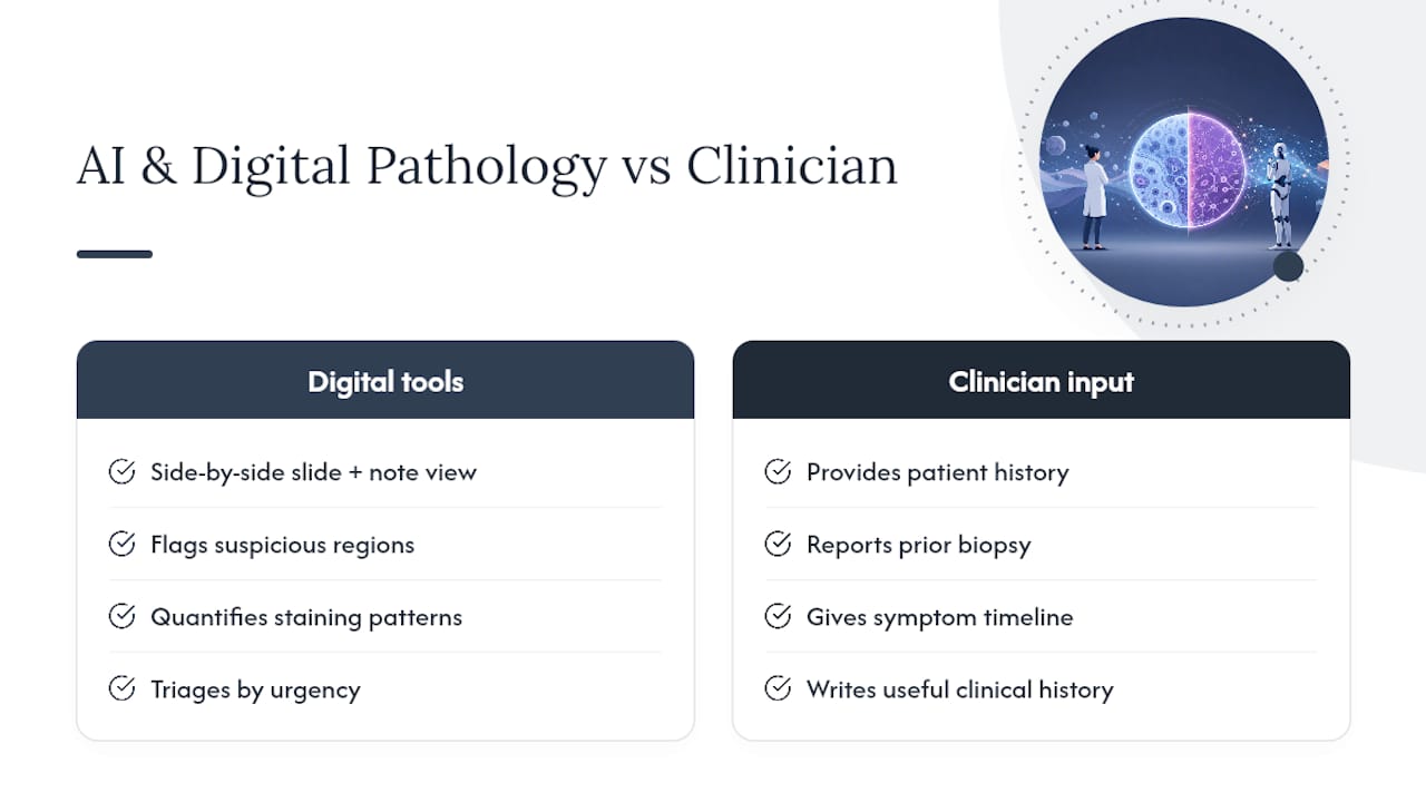
Digital pathology has changed what “sending the case to a pathologist” even means. Whole-slide imaging lets a pathologist pull up a scanned slide next to a clinician’s photo and clinical note on the same screen, instead of physically walking a slide tray between departments or waiting for a courier.
That proximity is the real value. AI tools built on top of digital pathology platforms are increasingly used to flag morphologic features for review, quantify staining patterns, or triage cases by urgency, but none of that replaces the correlation step itself. An algorithm can point at a suspicious region on a slide; it cannot know that the patient has a six-week history of rapid growth or a prior biopsy at the same site. That context still has to come from the clinician, and it still has to reach the pathologist before the read, not after.
Where digital tools genuinely help CPC is speed and access. A pathologist reviewing a difficult case can pull in a second opinion from a colleague in another building or another state without shipping a physical slide. Molecular and proteomic testing, increasingly guided by advances in peptide-based diagnostics, adds another data layer clinicians can request when morphology alone leaves the diagnosis genuinely uncertain. The technology expands what’s possible; it doesn’t substitute for a clinician who took the time to write a useful history.
How Can Departments Improve Communication Around CPC?
The biggest barrier to good CPC usually isn’t skill on either side of the microscope. It’s the absence of a reliable channel for clinicians and pathologists to talk to each other before a report is finalized, not after a discordant result forces the conversation.
Regular CPC conferences, even short ones, build the habit. A 15 or 20 minute weekly or biweekly session where a handful of hard cases get reviewed together, clinician and pathologist in the same room or on the same call, does more for diagnostic accuracy than any policy memo. It also teaches junior staff how each side actually thinks, which pays off long after the specific cases discussed are forgotten.
Outside of scheduled conferences, the fix is often as simple as making a direct line available. A pathologist who can call the ordering clinician with a quick question, rather than issuing a hedge-everything report because a detail is missing, produces better diagnoses. Departments that publish a named contact or a secure messaging channel for pathology questions see fewer of the “clinical correlation recommended” non-answers that leave both sides unsatisfied.
Cross-departmental case reviews, particularly for oncology, dermatology, and any specialty generating a high volume of biopsies, deserve a standing slot on someone’s calendar rather than an ad hoc effort that only happens when a case goes badly wrong.
How Do You Audit and Improve CPC Quality Over Time?
Quality assurance for CPC works best as a numbers exercise, not a vibe check. Tracking the rate at which cases get re-appraised, and how often that re-appraisal changes the diagnosis, gives a department a concrete baseline it can compare year over year.
A useful audit framework asks a short set of questions on a rolling basis: How often is clinical history missing or inadequate on requisitions? How often do pathologists request additional clinical information after the initial read? What share of discordant cases get resolved through a second opinion versus re-biopsy? Answering these regularly, even informally, surfaces systemic gaps that individual case reviews miss.
Documentation discipline is part of quality assurance too. A discordant case that gets resolved by phone call and never makes it into the chart teaches the department nothing the next time a similar case comes through. Recording the resolution, even briefly, turns each hard case into institutional knowledge rather than a one-off save.
Teaching CPC So It Actually Sticks
Run CPC rounds on short cases with one clear learning point each, whether that’s a diagnostic pitfall or the value of a single good photo. Have trainees rotate between presenting the clinical story and reading the pathology report critically, so both sides of the correlation become second nature.
— EIV Diagnostics
How EIV Diagnostics Supports Your CPC Workflow
EIV Diagnostics gives clinicians a faster path to the clinical context that makes correlation work, instead of a lab relationship where the specimen disappears into a queue and the report arrives with no conversation attached. Its molecular pathology and dermatopathology services are read by board-certified pathologists who factor in the clinical history you submit, not just the tissue on the slide.

Digital pathology workflows let you attach clinical or dermoscopic photos directly to the case, and mobile phlebotomy means a specimen or blood draw doesn’t have to wait for a clinic slot to open up. If a case needs a faster turnaround or a consultative second look, reach out through EIV Diagnostics before you finalize your requisition. To arrange testing, request a consult, or set up a mobile draw, visit EIV Diagnostics’ molecular pathology services and start the order with the clinical context that gives your pathologist something real to correlate against.
Sources
For deeper study, consult the teaching-focused CPC review, the pigmented skin tumor re-appraisal study, the neuroendocrine neoplasm collaboration analysis, and the UNC dermatology CPC handout.
This article is general information, not a substitute for advice from a qualified doctor. Consult a qualified healthcare professional about your own circumstances before acting on anything here.
- Clinico-pathological correlation: Teaching aspects, avenues, and advances
- Routine Clinical-Pathologic Correlation of Pigmented Skin Tumors Can Influence Patient Management
- The Clinician’s Role in Diagnostic Pathways and the Impact of Clinicopathological Collaboration on Outcomes in Patients with Neuroendocrine Neoplasms: Current Perspectives
FAQ
What Is a Clinicopathologic Correlation?
It is the process of matching a patient’s clinical history, exam findings, and test results against the gross and microscopic findings from a tissue sample to reach an accurate, patient-specific diagnosis.
What Does “Clinicopathological” Mean?
It describes relating a patient’s signs, symptoms, and lab results to what a pathologist observes on gross and histologic examination of tissue.
Is “Clinical Correlation Recommended” a Serious Finding?
It usually means the pathologist found something that can’t be fully classified from tissue alone and needs your clinical input to finalize the diagnosis, not that something is necessarily wrong.
What Does “Clinical Correlation” Mean on a Pathology Report?
It’s a note asking the ordering clinician to weigh the pathology findings against the patient’s actual presentation before treating the report as a final answer.
How Often Does CPC Actually Change a Diagnosis?
In one dermatopathology audit, re-appraisal was requested for 158 of 2,045 excised lesions, and 13 of those cases had their histologic diagnosis revised after correlation.