
By EIV Diagnostics · August 12, 2026
Fine Needle Aspiration: What Patients Need to Know
Discover what fine needle aspiration entails, its benefits, and how it aids in diagnosing various conditions quickly and safely.
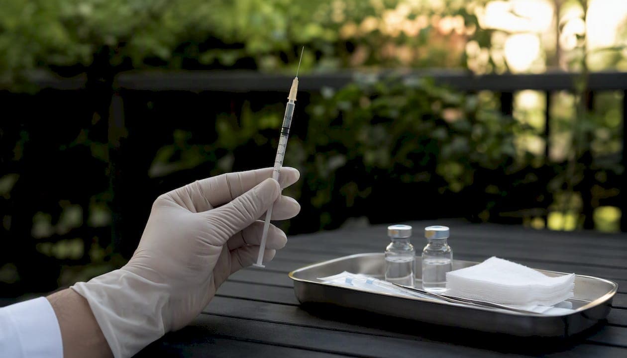
A fine needle aspiration (FNA) is a quick, minimally invasive procedure where a thin needle draws cells or fluid from a lump or mass for laboratory analysis. Most patients are in and out in under 30 minutes, with local anesthesia keeping discomfort minimal and routine cytology results typically ready within a few business days. According to StatPearls via the NIH, FNA is one of the most commonly used diagnostic tools for thyroid nodules, breast lumps, lymph nodes, and superficial soft-tissue masses, with a low overall complication rate. The Papanicolaou Society recommends it as part of a multidisciplinary, clinically guided approach rather than a standalone screening tool. Talk with your clinician about whether FNA or a different biopsy type is right for your specific situation.
Key Takeaways
FNA is a minimally invasive, quick diagnostic procedure with a strong track record when performed by experienced clinicians and read by qualified pathologists, and lab quality directly affects whether your result is diagnostic or not.
| Point | Details |
|---|---|
| What FNA is | A thin-needle procedure collecting cells or fluid for cytology, done in under 30 minutes with local anesthesia. |
| Diagnostic success rate | Experienced centers report approximately 92% diagnostic success; non-diagnostic samples may require a repeat procedure. |
| FNA vs core biopsy | FNA yields cytology; core-needle biopsy provides tissue architecture — the right choice depends on the diagnostic question. |
| Results timeline | Routine cytology takes roughly 3–4 business days; molecular ancillary tests take longer depending on the assay. |
| EIV Diagnostics | Offers cytology, molecular pathology, and digital pathology services to support FNA diagnostic workflows for clinicians and patients. |
Table of Contents
- What is a fine needle aspiration and when is it used?
- How does FNA differ from a core-needle or surgical biopsy?
- When do doctors recommend an FNA?
- How to prepare for your FNA appointment
- What happens during the procedure, step by step
- Recovery and aftercare after an FNA
- Risks and possible complications
- How FNA results are reported and what they mean
- Why the lab reading your FNA sample matters
- What patients commonly experience before, during, and after
- EIV Diagnostics supports FNA patients and providers with advanced lab services
- Sources
- FAQ
What is a fine needle aspiration and when is it used?
FNA removes cells or fluid from a suspicious mass using a very thin needle, typically 22–25 gauge, attached to a syringe. The sample goes to a pathology lab where a cytopathologist examines the cells under a microscope. That is the diagnostic use. There is also a therapeutic use: aspirating a cyst or seroma to relieve pressure or discomfort, even when no malignancy is suspected.
Common sites where FNA is performed include:
- Thyroid nodules — the most frequent application in the United States
- Breast lumps — both palpable and non-palpable masses
- Lymph nodes — especially enlarged or suspicious nodes in the neck, armpit, or groin
- Superficial soft-tissue lumps — lipomas, cysts, subcutaneous masses
- Deep or image-guided lesions — lung, liver, pancreas, or mediastinal masses sampled with CT or endoscopic ultrasound guidance
Imaging guidance, most often ultrasound, is used when the target is not easily felt by hand or sits near blood vessels or other vital structures. Ultrasound-guided FNA allows the clinician to watch the needle tip in real time, improving accuracy and safety. CT guidance is used for deeper lesions, and endoscopic ultrasound (EUS) is standard for pancreatic and mediastinal masses.
How does FNA differ from a core-needle or surgical biopsy?
The core difference is what each method retrieves. FNA collects individual cells for cytology. A core-needle biopsy (CNB) removes a small cylinder of tissue, preserving the architecture of how cells are arranged, which is often necessary for histologic diagnosis. Surgical or excisional biopsy removes the entire lesion or a large wedge of tissue.
| Feature | FNA | Core-Needle Biopsy | Excisional Biopsy |
|---|---|---|---|
| Needle/instrument size | Very thin (22–25 gauge) | Larger (core needle) | Surgical incision |
| Sample type | Cells (cytology) | Tissue core (histology) | Whole or partial lesion |
| Typical diagnostic yield | Variable; ~92% at experienced centers | Generally higher and more reproducible | Highest |
| Anesthesia | Local (often minimal) | Local | Local or general |
| Common uses | Thyroid, lymph nodes, cysts, quick cytology | Breast receptor testing, lymphoma workup, architecture-dependent diagnoses | Definitive removal or diagnosis |
| Recovery | Minutes to hours | Hours to a day | Days to weeks |
According to a PMC literature review on breast biopsy methods, FNA and CNB serve different diagnostic goals: FNA is faster and lower-risk, while CNB provides tissue architecture needed for receptor testing and histologic grading. The right choice depends on the diagnostic question, not a default preference.
FNA tends to be preferred for cystic lesions, lymph node cytology, thyroid nodules, and situations where anticoagulation makes a larger needle riskier. CNB is typically chosen when the pathologist needs to see tissue structure, such as distinguishing in-situ from invasive breast cancer, or when receptor and HER2 testing is required upfront.
Pro Tip: If your clinician suspects lymphoma or needs estrogen/progesterone receptor and HER2 status from a breast lesion before surgery, expect a core-needle biopsy rather than FNA. Those diagnoses require tissue architecture that a cytology sample cannot reliably provide.
When do doctors recommend an FNA?
Your clinician may refer you for this procedure in any of these situations:
- A new or recently changed lump that needs a tissue diagnosis
- An enlarged lymph node that hasn’t resolved after a few weeks
- A thyroid nodule flagged on ultrasound with suspicious features, per American Thyroid Association guidelines
- Drainage of a symptomatic cyst or abscess (selected cases)
- Sampling a known or suspected metastatic site for molecular marker testing
- Evaluation of a skin or subcutaneous mass before surgical planning
Imaging findings drive many of these referrals. A breast mass with a high BI-RADS category, a thyroid nodule with microcalcifications, or a lymph node with abnormal internal architecture on ultrasound can all trigger a biopsy recommendation. Multidisciplinary discussion between your radiologist, surgeon, and pathologist often shapes which biopsy type is ordered.
A few relative contraindications are worth knowing. FNA is generally avoided in actively infected fields, in patients with uncorrected severe coagulopathy, or when the result would not change clinical management. Your clinician will weigh these factors before scheduling the procedure.
How to prepare for your FNA appointment
Preparation for a superficial FNA is usually minimal, but a few steps matter.
- Medications: Tell your clinician about all anticoagulants (warfarin, rivaroxaban, apixaban), antiplatelet agents (aspirin, clopidogrel), and herbal supplements (fish oil, vitamin E, ginkgo) that can increase bleeding risk. Your team will decide whether to hold them or proceed with extra precautions.
- Fasting: Most superficial FNAs require no fasting. If your procedure involves sedation (such as endoscopic ultrasound-guided FNA), your facility will give you specific fasting and medication instructions — follow those exactly.
- What to bring: Referral slip, photo ID, insurance card, a complete medication list, any prior imaging (ultrasound, CT, MRI reports or discs), and previous pathology or biopsy results if available.
- Anti-anxiety medications: Some facilities ask patients not to take sedating medications before the procedure. Confirm this with your clinic in advance rather than assuming it is fine.
- Support: Bring a friend or family member if you want someone with you. Most patients drive themselves home afterward, but having company can help if you are anxious.
According to StatPearls, many superficial palpable FNAs require minimal to no patient preparation. The more involved the guidance method (CT, endoscopic), the more preparation your facility will require.
What happens during the procedure, step by step
- Check-in and consent. You review and sign an informed consent form. The clinician explains the procedure, risks, and alternatives.
- Positioning and palpation. You are positioned so the target area is accessible. The clinician feels the lump or reviews imaging to plan the approach.
- Skin preparation. The skin over the site is cleaned with antiseptic.
- Local anesthetic (if used). A small amount of lidocaine may be injected to numb the skin. Interestingly, StatPearls notes that the anesthetic injection itself can sometimes cause more discomfort than the fine needle, so some clinicians skip it for very superficial passes.
- Needle insertion and aspiration. The thin needle enters the mass. The clinician moves it back and forth in short strokes while applying gentle suction. Each individual pass lasts roughly 10–15 seconds. Multiple passes (typically 2–4) are made to collect enough material.
- Ultrasound guidance (when applicable). For non-palpable or deep lesions, real-time ultrasound guidance lets the radiologist or cytopathologist watch the needle tip throughout the procedure. Even palpable lumps are often sampled under ultrasound to maximize yield and reduce the need for repeat procedures.
- Slide preparation or specimen collection. The aspirated material is expressed onto glass slides or into a liquid medium immediately. Rapid on-site evaluation (ROSE) means a cytopathologist reviews the slides right there to confirm adequate cellularity before you leave.
- Brief observation. Pressure is applied to the site. You rest for a few minutes while the team confirms the sample looks adequate.
The total appointment runs 20–30 minutes at most experienced centers. The needle-in-tissue time is a fraction of that.
On diagnostic adequacy: At experienced centers, FNA achieves a diagnostic success rate of approximately 92%, with non-diagnostic rates near 1% in some series. Rates vary by operator skill, lesion type, and whether on-site evaluation is available. A non-diagnostic result does not mean something is wrong — it means the sample did not contain enough evaluable cells, and a repeat procedure or alternative biopsy is typically the next step.
Recovery and aftercare after an FNA
Most patients feel mild soreness at the puncture site for 24–48 hours. Bruising is common and usually resolves within a week.
- Ice: Apply a cold pack wrapped in cloth for 10–15 minutes at a time during the first few hours to reduce swelling and bruising.
- Pain relief: Acetaminophen (Tylenol) works well for most patients. Avoid aspirin and NSAIDs like ibuprofen for the first 24 hours unless your clinician says otherwise, since they can increase bleeding.
- Activity: Most patients return to normal activities the same day. Avoid strenuous exercise or heavy lifting for 24 hours if the site is tender.
- Wound care: Keep the puncture site clean and dry. A small bandage is usually all that is needed.
- Blood thinners: Resume anticoagulants only when your clinician confirms it is safe — do not restart them on your own schedule.
Pro Tip: If visible bruising bothers you, arnica gel applied to the skin (not the puncture itself) can help it fade faster. Most bruising peaks around day 2–3 and clears by day 7–10.
Call your clinic or seek urgent care if you notice:
- Increasing pain or swelling after the first 24 hours
- Fever above 100.4°F
- Heavy or uncontrolled bleeding at the site
- Redness, warmth, or discharge suggesting infection
- Shortness of breath or chest pain (relevant for neck or chest FNAs)
Risks and possible complications
FNA is considered a low-risk procedure, but no needle procedure is entirely without risk.
- Bruising and mild pain — the most common outcomes, affecting many patients to some degree; typically self-limiting
- Bleeding — minor oozing is normal; significant hematoma is rare
- Infection — uncommon when standard sterile technique is used
- Damage to nearby structures — rare; organ-specific examples include pneumothorax from a lung or neck FNA, or hoarseness from a thyroid FNA near the recurrent laryngeal nerve
- Non-diagnostic sample — not a complication in the traditional sense, but a recognized limitation; it occurs more often with certain lesion types and less experienced operators
- Needle-tract seeding — theoretically possible for certain tumors (notably some liver and pancreatic lesions), though the clinical risk is considered very low with modern thin needles and is far outweighed by the diagnostic benefit in most situations
- Vasovagal reaction — some patients feel lightheaded or faint briefly; lying down during the procedure reduces this risk
Diagnostic performance varies meaningfully by organ and operator. A PMC review of breast biopsy literature found FNA sensitivity ranging from 35% to 95% in older breast studies, while CNB tends to deliver higher and more reproducible sensitivity in many settings. For thyroid nodules, FNA remains the standard first-line diagnostic tool with well-established performance when performed by experienced hands. Contact your clinic if any symptom feels unusual or worsening — most complications, when they occur, are manageable early.
How FNA results are reported and what they mean
Routine cytology results typically come back in 3–4 business days. Rapid on-site evaluation gives a preliminary read the same day. Specialized molecular tests take longer, depending on the lab and the specific assay.
| Result Category | What It Means | Typical Next Step |
|---|---|---|
| Benign | Cells appear normal; no malignancy identified | Monitoring, imaging follow-up, or no further action |
| Malignant | Cancer cells identified | Surgical planning, oncology referral, staging workup |
| Suspicious | Features concerning but not definitive | Core-needle biopsy or surgical excision |
| Atypia / Indeterminate | Abnormal cells present but classification uncertain | Repeat FNA, molecular testing, or CNB |
| Non-diagnostic | Insufficient cells for evaluation | Repeat FNA or alternative biopsy |
The Bethesda System is the standard reporting framework for thyroid FNA cytology, using six categories (I through VI) that map directly to malignancy risk and recommended management. Breast FNA uses a similar five-category system. Your pathology report will typically include the category, a description of the cells seen, and sometimes a recommendation for next steps.
Pro Tip: When your results arrive, ask your clinician three specific questions: How confident is the pathologist in this result? Is molecular testing available to refine the diagnosis? And if the result is indeterminate, what is the recommended next step and timeline?
For molecular diagnostic testing on FNA material, such as BRAF mutation testing for thyroid nodules or next-generation sequencing for metastatic lesions, turnaround times vary by assay and lab. Ask upfront whether your sample will be sent for ancillary testing and how that affects your overall timeline.
Why the lab reading your FNA sample matters
The clinician who takes the sample and the pathologist who reads it are equally important to getting a useful result. A technically adequate aspirate can still yield a non-diagnostic report if the cytopathologist lacks experience with that tissue type, and a skilled cytopathologist cannot compensate for a poorly collected sample.
Key quality signals to look for in the lab processing your FNA:
- Board-certified cytopathologists or pathologists with documented experience in cytology
- Rapid on-site evaluation (ROSE) capability, which allows real-time adequacy assessment and reduces repeat procedures
- Molecular pathology capability for ancillary testing (BRAF, KRAS, next-generation sequencing panels) directly on FNA material
- CAP and CLIA accreditation confirming the lab meets federal quality and proficiency standards
- Digital pathology access enabling remote expert review or second opinions on difficult cases
StatPearls is direct on this point: outcomes improve when the clinician obtaining the sample and the interpreting cytopathologist collaborate, and on-site evaluation with board-certified pathologists reduces non-diagnostic sampling while speeding decision-making.
The workflow matters too. Once aspirated, your sample moves through slide preparation, cytology review, and potentially ancillary testing (immunohistochemistry, molecular assays). Each handoff is a point where quality controls either protect or compromise your result. A lab with integrated molecular pathology can run additional tests on the same FNA material without requiring a second procedure.
What patients commonly experience before, during, and after
Anxiety before the appointment is normal and nearly universal. Most patients describe the actual procedure as far less uncomfortable than they expected. The needle is thin, the passes are brief, and the local anesthetic, when used, handles most of the sharpness.
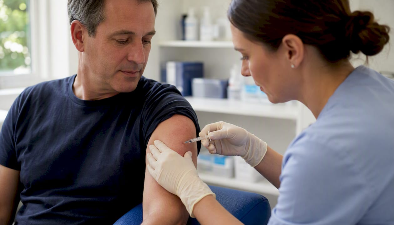
During the procedure, patients often notice pressure more than pain. Some feel a brief sting with the anesthetic injection. A few experience a vasovagal response (lightheadedness) and recover quickly once they lie flat.
Afterward, the most common report is mild soreness for a day or two, manageable with acetaminophen. Most people return to work or normal activities the same afternoon. The harder part, for many, is the wait for results. Knowing that routine cytology typically takes 3–4 business days, and that a non-diagnostic result is a technical outcome rather than a verdict, helps reduce that anxiety.
Bring a friend if you want company. Write down your questions before the appointment. Ask the clinician to explain what they are doing as they go — most are happy to narrate the procedure in real time.
EIV Diagnostics supports FNA patients and providers with advanced lab services

When your FNA sample leaves the procedure room, where it goes next shapes everything about your result. EIV Diagnostics is an independent pathology laboratory in Fresno, CA, staffed by board-certified pathologists and built to handle the full diagnostic workflow that FNA material demands.
EIV Diagnostics offers cytology services for FNA specimens, molecular pathology for ancillary testing on aspirated material, and digital pathology for remote expert review and second opinions. The lab works with healthcare providers ordering diagnostic services and with patients who want to understand their options directly. EIV Diagnostics does not perform the clinical FNA procedure itself — that is your clinician’s role. The lab’s job is to deliver accurate, timely diagnostic reports and advanced testing that inform what happens next.
Clinicians can contact EIV Diagnostics for specimen-handling guidance and test ordering. Patients with questions about their pathology results or available molecular tests are welcome to reach out as well.
Sources
The following sources informed this article and are worth bookmarking for deeper reading:
- Fine Needle Aspiration - StatPearls - NCBI Bookshelf - NIH
- Fine-needle versus core-needle biopsy – which one to choose in preoperative assessment of focal lesions in the breasts? Literature review - PMC
- Fine Needle Aspiration
- Ultrasound Guided Fine Needle Aspiration | Radiology
- Ultrasound Guided Fine Needle Aspiration
For questions specific to your case, contact your clinician or the laboratory processing your sample. These sources provide general information and do not replace individualized medical advice.
This article is for general informational purposes only and is not a substitute for professional medical advice. Always confirm current clinical recommendations with your healthcare provider or a qualified specialist.
FAQ
How painful is a fine needle aspiration?
Most patients describe FNA as mildly uncomfortable rather than painful. The needle is very thin, individual passes last roughly 10–15 seconds, and local anesthetic is commonly used to numb the skin beforehand.
What is the difference between a biopsy and a fine needle aspiration?
FNA is a type of biopsy that collects individual cells using a thin needle for cytology review. A core-needle biopsy uses a larger needle to remove a tissue cylinder, providing the architectural detail needed for histologic diagnosis and receptor testing.
Do they put you to sleep for a fine needle aspiration?
No. Most FNAs use only local anesthesia to numb the skin. General or IV sedation is reserved for endoscopic ultrasound-guided procedures, and your facility will give you specific preparation instructions if sedation is planned.
How long does it take to recover from a fine needle aspiration?
Recovery is typically same-day. Most patients experience mild soreness and possible bruising for 24–48 hours, manageable with acetaminophen, and return to normal activities within hours of the procedure.
Recommended
- Dermatopathology Services: What Patients and Providers Need to Know | EIV Diagnostics | EIV Diagnostics
- Molecular Diagnostic Testing: What Patients and Clinicians Need to Know | EIV Diagnostics | EIV Diagnostics
- Clinical Pathology Labs: What Patients Need to Know | EIV Diagnostics | EIV Diagnostics
- Cytology Testing in Tulare County: What You Need to Know | EIV Diagnostics | EIV Diagnostics