
By EIV Diagnostics · September 24, 2026
Digital Pathology: 98.3% Concordance and When Labs Still Need Glass
Operational guide for lab leaders explaining evidence-backed benefits, real cost and turnaround tradeoffs, and a practical validation checklist with EIV...
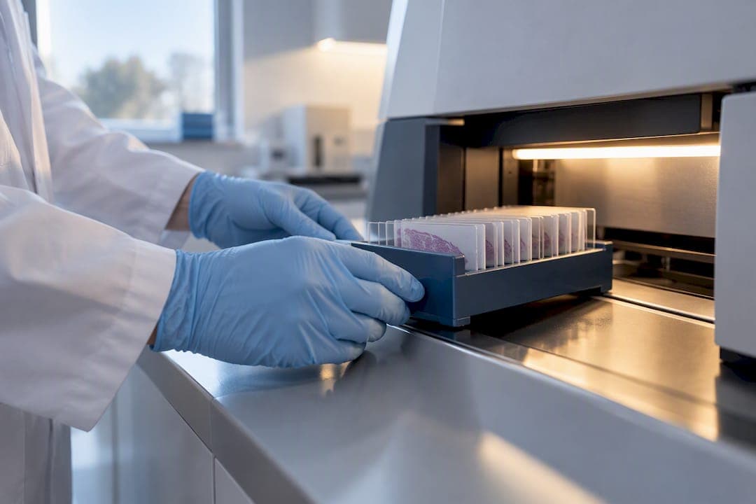
Digital pathology cuts turnaround time, opens the door to remote subspecialty consults, and creates a searchable slide archive that also feeds AI-assisted image analysis. It matches light microscopy in the vast majority of cases, but grading calls and small objects still trip up digital review often enough that labs need local validation, a trained team, and real infrastructure before they cut over. Some labs treat the validation step as the actual product, not paperwork.
TL;DR:
- Digital pathology significantly reduces turnaround times by enabling case routing and remote review, especially in multi-site and subspecialty settings.
- Its diagnostic concordance with light microscopy averages 98.3%, but grading and small object detection still require fallback protocols and validation.
- Cost savings primarily stem from fewer courier trips and quicker case access, but high upfront investments and ongoing IT costs can extend payback periods for low-volume labs.
- AI tools excel in triage, quantification, and marker scoring when validated locally and used as decision-support, not standalone diagnostics.
- Implementation success depends on thorough validation, proper infrastructure, and staged rollout with staff training and QC procedures.
Table of Contents
- What Are the Operational Benefits of Digital Pathology?
- How Accurate Is Digital Pathology Compared to Light Microscopy?
- Does Digital Pathology Actually Save Time and Money?
- Where Does AI Add Real Value in Digital Pathology?
- What Are the Downsides and Risks of Digital Pathology?
- How Do You Implement and Validate a Digital Pathology System?
- How EIV Diagnostics Approaches Digital Pathology in Practice
- When Is Digital Pathology the Right Strategic Move?
- Ready to Explore Digital Pathology for Your Lab?
- Sources
- FAQ
What Are the Operational Benefits of Digital Pathology?
The biggest shift isn’t diagnostic. It’s logistical. Once slides exist as whole-slide images instead of physical glass, a lab stops shipping trays between sites and starts routing cases to whoever is available to read them, regardless of location.
That single change ripples through the whole department. A pathologist covering three satellite clinics can sign out cases from one screen instead of driving between buildings. A rare tumor board consult that used to take a week of courier time now happens the same afternoon over a shared viewer. Retired or part-time subspecialists can keep contributing without commuting to a hub lab.
Digital pathology also strengthens research and training. Archived cases become searchable teaching sets, and multicenter clinical trials benefit from centralized, blinded review that keeps scoring consistent across sites, something paper requisitions and physical slide shuffling never managed reliably.
None of this happens automatically. Realizing these gains requires a working stack: a validated scanner, image storage with enough capacity for whole-slide files, an image management system, viewer software your pathologists actually like using, and integration with your existing LIS or EHR.
What this changes day to day:
- Cases move between reviewers in minutes, not days, cutting courier dependency almost entirely.
- Subspecialty opinions arrive without shipping a single slide.
- Archived studies become instantly searchable for teaching, audits, or trial recruitment.
- Staffing gets more flexible since sign-out no longer requires physical presence.
Pro Tip: Before buying a scanner, map your current slide-to-signout workflow on paper. The bottleneck is rarely the microscope. It’s usually the handoff points between grossing, cutting, and delivery to the pathologist’s desk, and digitizing those steps first often produces faster wins than the imaging hardware itself.
How Accurate Is Digital Pathology Compared to Light Microscopy?
A 2020 systematic review and meta-analysis covering 25 studies and 10,410 histology samples found 98.3% overall concordance between digital pathology and conventional light microscopy, with a 95% confidence interval of 97.4% to 98.9%.
Most discordances in that analysis broke down into three categories: 57% involved nuclear atypia or grading and dysplasia calls, 26% were generally challenging diagnoses regardless of viewing method, and 16% involved small objects that are easy to miss on a screen but visible under a microscope’s fine focus control.
Grading disputes and tiny structures, think mitotic figures, microorganisms, or small calcifications, are where digital review earns its reputation for caution. The likely explanation is straightforward: whole-slide scanners typically capture a single focal plane, while glass slides let a pathologist rack through z-axis depth in real time.
What this means for your protocols:
- Keep glass-slide review accessible for grading-heavy specimens like breast or prostate biopsies.
- Build explicit escalation rules for any case involving small-object detection.
- Don’t treat 98.3% concordance as a green light to skip a fallback pathway entirely.
Does Digital Pathology Actually Save Time and Money?
A departmental deployment study in Spain gives one of the clearer real-world signals here. Across 11,922 cases analyzed, mean turnaround time dropped significantly after switching to digital pathology, resulting in a notable reduction. Pathologist workload fell substantially on average during the first year of adoption.
Those numbers came from one specific lab with its own case mix, staffing, and scanner setup, so treat them as a directional benchmark rather than a guarantee.
Where the savings actually come from:
- Fewer courier trips and less slide handling between departments.
- Faster access to archived cases means fewer repeat cuts and fewer delays waiting on physical slide retrieval.
- Remote consultation capacity increases referral and second-opinion volume without adding headcount.
Where the costs bite back:
- Scanner hardware and storage infrastructure require upfront capital.
- Informatics staffing and IT integration are recurring costs, not one-time purchases.
- Payback timelines stretch out for smaller labs with lower case volume, since fixed costs get spread across fewer studies.
Scale matters more than almost any other variable. A high-volume lab reusing its archive for research or AI training will recoup costs faster than a low-volume practice running the same hardware for diagnostic work alone.
Where Does AI Add Real Value in Digital Pathology?
AI-enabled image analysis is where digital pathology stops being a viewing tool and starts becoming a decision-support tool, but the gap between marketing claims and validated performance is wide. The technologies that actually work today tend to be narrow and well-defined rather than broadly diagnostic.
Realistic, currently useful applications include triage heatmaps that flag suspicious regions for prioritized review, automated mitotic counts for grading workflows, and quantitative scoring for biomarkers like PD-L1 or HER2, tasks that are tedious and variable when done manually.
What labs need before trusting an algorithm:
- Local validation against your own case mix, not just the vendor’s published performance.
- A clearly defined role for the tool: triage assistance versus diagnostic confirmation are very different risk profiles.
- Ongoing monitoring, since AI performance tends to drift as case populations shift over time.
The FDA maintains an AI-enabled medical device list showing devices cleared for specific intended uses. A clearance for one application doesn’t transfer to another use case, so confirming labeling before deployment matters as much as the clearance itself.
Pro Tip: Ask any AI vendor for their validation dataset’s demographic and specimen breakdown before you sign anything. An algorithm trained mostly on one tissue type or population can underperform badly on your actual case mix, and you won’t find that out until after go-live.
What Are the Downsides and Risks of Digital Pathology?
Every lab that has gone digital has hit at least one of these friction points. Planning around them upfront beats discovering them mid-rollout.
- Z-plane and focus limits. Most scanners capture one focal plane, so structures visible under manual focus adjustment can be missed on a flat digital image.
- Small-object visibility. Parasites, microorganisms, and fine calcifications are the recurring blind spot noted across discordance studies.
- Display and calibration drift. Monitors need regular calibration; an uncalibrated screen changes color and contrast enough to affect subtle calls.
- Storage and network strain. Whole-slide images run into gigabytes each, and undersized infrastructure creates lag that frustrates pathologists fast.
- Staff resistance. Pathologists trained exclusively on glass slides often need real time and structured practice to trust a screen for primary diagnosis.
- Missing QC and SOPs. Rolling out scanners without new quality-control checkpoints and documented escalation rules is the single most common early-stage mistake.
Staged rollouts, mandatory glass-slide fallback for flagged categories, and dedicated training time solve most of this before it becomes a crisis.
How Do You Implement and Validate a Digital Pathology System?
The College of American Pathologists requires laboratories to validate whole-slide imaging systems locally before clinical use. Vendor performance claims don’t substitute for testing your own specimen mix, staff, and workflow.
Technical stack to confirm before go-live:
- Validated scanner and calibrated displays matched to your case volume.
- Image management system with adequate storage and backup redundancy.
- Viewer software integrated with your LIS or EHR.
- Cybersecurity protocols covering image transfer and storage.
Rollout steps that hold up in practice:
- Run a pilot on a limited case type before expanding scope.
- Design a validation study sized to your specimen mix, not a generic template.
- Write SOPs covering escalation, QC checkpoints, and data retention.
- Train every pathologist who will read digitally, not just early adopters.
| Implementation Stage | Primary Focus | Key Reference |
|---|---|---|
| Pilot | Limited case type, controlled comparison to glass slides | CAP validation guidance |
| Validation study | Sample sized to your own specimen mix | CAP validation guidance |
| Rollout | SOPs, training, QC checkpoints | Internal quality program |
| Monitoring | Ongoing concordance checks, AI drift review | FDA device labeling |
How EIV Diagnostics Approaches Digital Pathology in Practice
EIV Diagnostics runs digital pathology alongside molecular pathology, dermatopathology, and confocal microscopy, which means digitized slides don’t sit in isolation. A dermatopathology case flagged during digital review can move straight into molecular testing without a new sample request.
Board-certified pathologists handle sign-out, and mobile phlebotomy covers the sample-logistics side for providers who need draws completed away from a central lab.
What this looks like for a referring provider or lab partner:
- Digital slide review paired with subspecialty sign-out rather than a generic read.
- A path from digital pathology into molecular pharmacogenomic testing when a case calls for it.
- Mobile phlebotomy support for practices without in-house draw capacity.
Labs weighing a pilot or validation partnership can start with a direct conversation about case mix and volume before committing to hardware.
When Is Digital Pathology the Right Strategic Move?
Digital pathology makes strategic sense when volume, subspecialty demand, or multi-site coverage justify the infrastructure. A single-site lab with steady case volume and no consult backlog may not need it yet. A network running multiple locations, chasing subspecialty expertise, or supporting clinical trials almost always benefits.

The most common premature mistake is buying scanners before building the informatics and validation plan around them. Hardware is the easy purchase. Staffing, storage, and CAP-aligned validation are the parts that actually determine whether the investment pays off.
Track turnaround time, pathologist workload, and consult volume for the first year. Those three numbers tell you faster than any vendor pitch whether the switch is working.
— EIV Diagnostics
Ready to Explore Digital Pathology for Your Lab?
EIV Diagnostics runs digital pathology as one piece of a connected diagnostic operation, not a standalone scanner purchase. A case reviewed digitally can move directly into molecular pathology or dermatopathology workup without a new specimen request, and board-certified pathologists handle sign-out on every case.

For providers building or evaluating a digital pathology workflow, building or evaluating a digital pathology workflow may benefit from a practical starting point involving a conversation about case mix, volume, and validation needs before any hardware decision is made. Mobile phlebotomy is also available for practices that need draws completed off-site; current prices are available on the provider’s website. Reach out through the digital pathology services page to discuss a pilot or validation partnership tailored to your lab’s actual specimen mix.
Sources
- Diagnostic concordance and discordance in digital pathology: a systematic review and meta-analysis
- Validating Whole Slide Imaging For Diagnostic Purposes In Pathology - CAP
- AI-Enabled Medical Device List — FDA
This article is general information, not a substitute for advice from a qualified doctor. Consult a qualified healthcare professional about your own circumstances before acting on anything here.
FAQ
What Are the Downsides of Digital Pathology?
The main downsides are technical limits around focus depth and small-object detection, plus the operational cost of staff training, storage infrastructure, and new quality-control procedures. Grading-heavy specimens and cases involving tiny structures like microorganisms carry the highest risk of discordance, which is why a glass-slide fallback still belongs in most protocols.
Is Digital Pathology FDA Approved?
Whole-slide imaging systems and specific AI-enabled pathology devices can receive FDA clearance, but clearance applies to a defined intended use, not to digital pathology as a general category. The FDA’s AI-enabled device list shows exactly which devices are cleared and for what specific application.
Is AI Going to Replace Pathologists?
No. Current evidence supports AI as a triage and quantification aid, flagging regions of interest or scoring biomarkers, not as a replacement for pathologist judgment. Every algorithm still needs role-specific validation before a lab can trust its output for diagnostic decisions.
What Does a Digital Pathologist Do?
A digital pathologist is a qualified pathologist who interprets whole-slide images using validated scanners, calibrated displays, and viewer software, correlating those images with clinical and lab data to issue a diagnostic report. The role also covers remote consultation, quality control, case triage, and supervised use of image-analysis tools.
Does EIV Diagnostics Offer Digital Pathology Services?
Yes. EIV Diagnostics provides digital pathology alongside molecular pathology, dermatopathology, and confocal microscopy, with board-certified pathologists handling sign-out. Pricing for most services is available on request through the site; mobile phlebotomy is listed from $65 per visit.