
August 21, 2026
Confocal Microscopy for Melanoma: Accuracy, Uses, and Limits
Discover how confocal microscopy enhances melanoma diagnosis, improving accuracy and reducing the need for invasive biopsies.
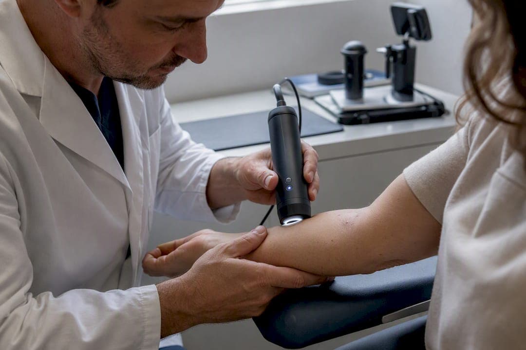
Reflectance confocal microscopy (RCM) is a validated adjunct to dermoscopy that improves melanoma diagnosis without replacing biopsy or histopathology. It does not stand in for a scalpel, but it does change how often that scalpel gets used.
The evidence is now deep enough to state numbers with confidence. Pooled analyses put RCM’s sensitivity around 90-93% and specificity between 70% and 82%, depending heavily on which patients get imaged. A randomized clinical trial published in JAMA Dermatology found that adding RCM to standard evaluation cut the number needed to excise (NNE) by roughly 43%, from 5.3 down to 3.0, while positive predictive value nearly doubled, from under 20% to above 30%.
What that means in practice:
- RCM earns its place on equivocal pigmented lesions after dermoscopy has already been done, not before.
- It should never override a strongly suspicious clinical or dermoscopic finding, especially on nodular or ulcerated lesions.
- Accuracy depends on operator experience and lesion selection. Consecutive lesion series score better on specificity than cohorts of preselected suspicious lesions.
- Cochrane-style systematic review data and a randomized trial (not just retrospective case series) back these numbers, which is rare for a dermatologic imaging tool.
The takeaway for a working clinic: RCM shifts the biopsy threshold intelligently. It does not eliminate biopsy. Anyone selling it as a replacement for histopathology is misreading the literature.
Key Takeaways
RCM improves melanoma detection accuracy and cuts unnecessary excisions, but it works only as an adjunct to dermoscopy and histopathologic confirmation, never a replacement for either.
| Point | Details |
|---|---|
| Accuracy is context-dependent | Pooled sensitivity runs 90 to 93% and specificity 70 to 82%, shifting with lesion selection and operator experience. |
| RCM cuts excisions, not detection | The JAMA trial found a 43% NNE reduction without lowering melanoma detection rates. |
| Know the failure modes | Ulceration, crust, hyperkeratosis, and acral skin block reliable imaging and can hide true positives. |
| Scoring adds consistency | The Pellacani score and similar algorithms give reproducible thresholds but must pair with clinical judgment. |
| EIV Diagnostics closes the loop | EIV Diagnostics provides dermatopathology, histopathology, and digital pathology to confirm RCM-flagged lesions with board-certified reporting. |
Table of Contents
- What Is Confocal Microscopy for Melanoma and How Does It Work?
- How Accurate Is Confocal Microscopy for Detecting Melanoma?
- When Should Clinicians Use RCM for a Suspicious Mole?
- What Does Melanoma Look Like Under Confocal Microscopy?
- What Are the Limitations of Confocal Microscopy for Melanoma?
- How Do You Fit RCM Into a Dermatology Clinic Workflow?
- What Scoring Systems Help Interpret Confocal Images?
- How Should RCM Findings Be Documented and Reviewed?
- A Practical Checklist for Using Confocal Microscopy on a Suspicious Lesion
- How EIV Diagnostics Supports RCM-Based Melanoma Workups
- Where RCM Fits in the Bigger Picture of Melanoma Care
- Referring Suspicious Lesions for Pathology Confirmation
- Key Studies and Reviews Worth Reading
- Sources
- FAQ
What Is Confocal Microscopy for Melanoma and How Does It Work?
Reflectance confocal microscopy uses near-infrared light, typically around 830 nanometers, to generate horizontal optical sections of skin in real time, without cutting anything. Melanin acts as a natural reflector, so pigmented structures light up against surrounding tissue with a contrast that dermoscopy simply cannot match at the cellular level.
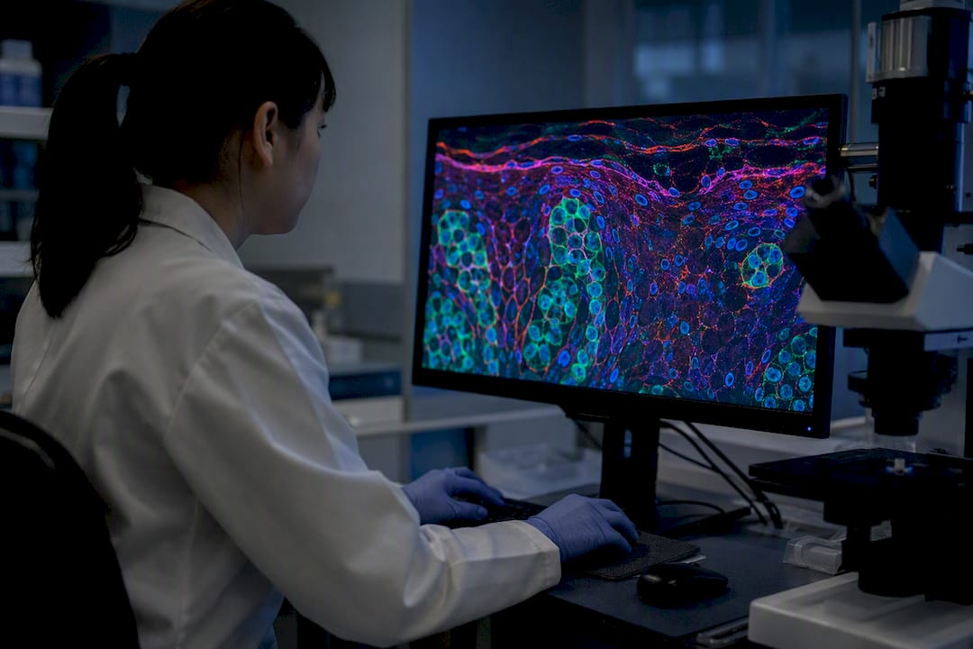
The imaging reaches the papillary and upper reticular dermis, with lateral resolution near 1 micrometer, close enough to histologic detail that trained readers can identify individual keratinocytes, melanocytes, and dendritic cells. That is the whole appeal: you get near-histologic information on a living patient, in the exam room, before deciding whether to cut.
Static units versus handheld probes
Two device categories dominate clinical use, and they solve different problems.
- Static, stage-mounted systems like the VivaScope 1500 use a wide-field mosaic approach, stitching together many individual fields into a large map of a lesion. This makes them well suited to lesion mapping, margin assessment before excision, and documentation of large or irregular pigmented patches such as suspected lentigo maligna.
- Handheld probes like the VivaScope 3000 trade mosaic coverage for speed and maneuverability. They are built for rapid point-of-care screening, awkward anatomic locations (near the eye, ear, or nose), and quick second-look confirmation during a routine skin exam.
Neither device is objectively better. A static mosaic unit is the right call when you need a reproducible, archivable map of a 15mm lesion on the back. A handheld probe wins when you need a 90-second answer on a small lesion on the nose during a busy clinic day.
Pro Tip: Don’t image a lesion that’s freshly cleaned with alcohol or still wet from an antiseptic wipe. Surface moisture and residue scatter the near-infrared beam and produce a hazy, low-contrast image that mimics genuine architectural disorder.
The physics also explains RCM’s failure modes before you ever pick up a probe. Melanin reflects light beautifully, which is why RCM excels on pigmented lesions. But hyperkeratosis, crust, and ulceration scatter and absorb that same light before it reaches the dermis, which is why heavily keratotic or bleeding lesions produce unreadable images no matter how skilled the operator is. Keep that limitation in mind: it resurfaces later as one of the most consequential pitfalls in melanoma imaging.
How Accurate Is Confocal Microscopy for Detecting Melanoma?
The honest answer is: quite good, but the number you quote depends entirely on which patients were imaged.
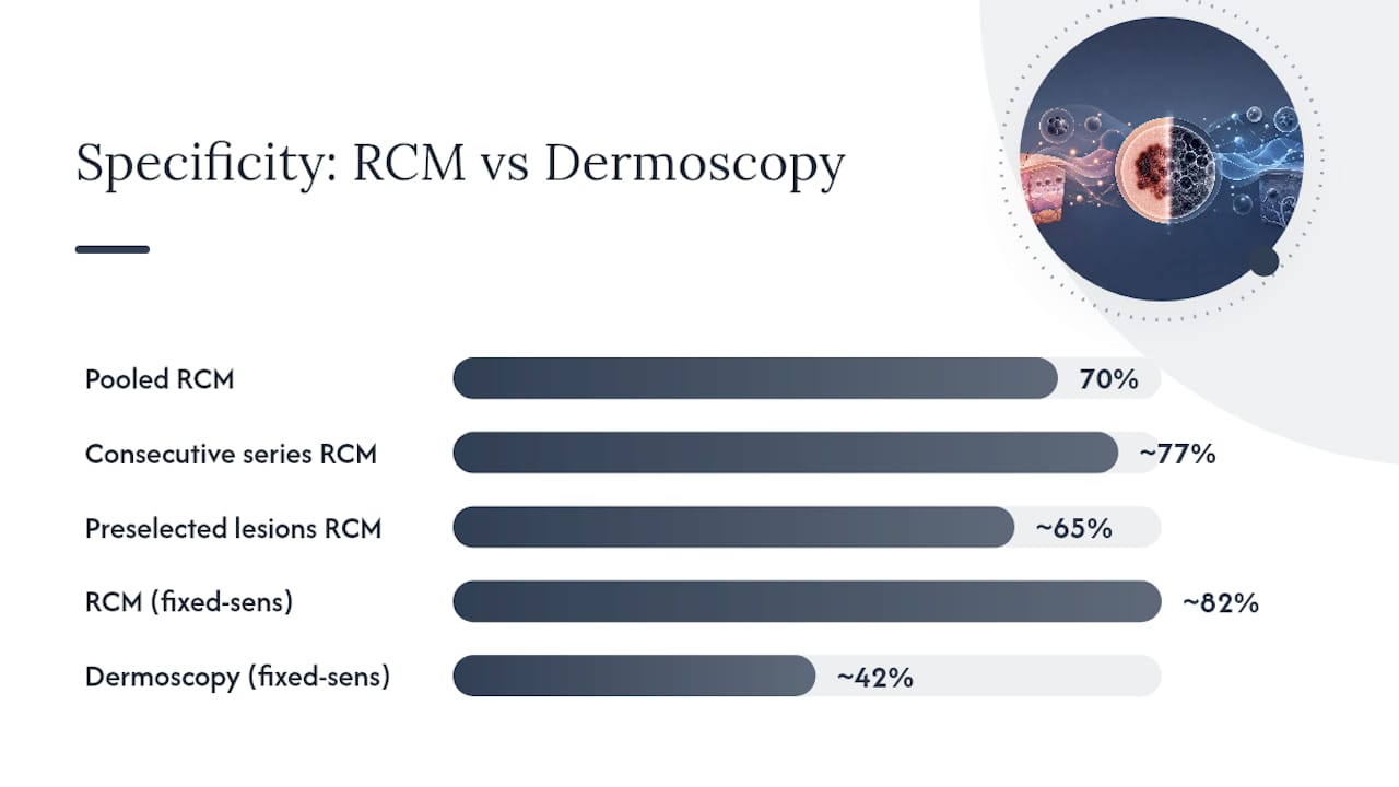
A meta-analysis covering 32 studies and 7,352 lesions found pooled sensitivity of 92% (95% CI: 0.91-0.93) and pooled specificity of 70% (95% CI: 0.69-0.71) for RCM in diagnosing malignant melanoma. That headline number hides real variation, though. Specificity climbed to around 77% in consecutive lesion series, where clinicians imaged everything that came through the door, and dropped to roughly 65% in cohorts built from highly suspicious, preselected lesions.
That gap matters more than it looks. A test performs differently depending on the base rate of disease in front of it, and RCM is no exception. Feed it a mix of ordinary nevi and the occasional melanoma, and specificity looks strong. Feed it a stack of lesions already flagged as worrisome by dermoscopy, and specificity drops because you have concentrated the ambiguous cases.
A separate systematic review reinforced the point from a different angle: RCM outperformed dermoscopy directly, with an estimated specificity near 82% for RCM against roughly 42% for dermoscopy when sensitivity was held fixed. That is the clearest evidence that RCM adds real diagnostic value on top of dermoscopy rather than simply duplicating it.
| Study context | Sensitivity | Specificity | Source |
|---|---|---|---|
| Pooled meta-analysis, 32 studies | 92% | 70% | Wiley systematic review |
| Consecutive lesion series | Similar to pooled | ~77% | Wiley systematic review |
| Highly suspicious, preselected lesions | Similar to pooled | ~65% | Wiley systematic review |
| RCM vs. dermoscopy (fixed sensitivity) | Comparable | 82% (RCM) vs. 42% (dermoscopy) | PMC systematic review |
The randomized trial evidence is where things get practically interesting. In a trial of 3,165 patients with suspect lesions, adjunctive RCM reduced the number needed to excise from 5.3 down to 3.0, a 43.2% reduction, while positive predictive value rose from 18.9% to 33.3%. Melanoma detection rates held steady between groups, meaning the accuracy gain came from cutting unnecessary excisions of benign lesions, not from missing more cancers. The few delayed diagnoses that did occur in the RCM arm were thin melanomas, not aggressive or late-stage disease.
A few caveats keep this evidence honest rather than promotional.
- Heterogeneity across studies is real. Pooled numbers smooth over differences in operator skill, device type, and lesion population that individual clinics will not experience uniformly.
- Selection bias runs through most cohort studies, since lesions chosen for RCM often skip the most obviously benign cases and the most obviously malignant nodular ones, both of which are harder for RCM to characterize well.
- Operator experience correlates strongly with accuracy. The JAMA trial found physicians’ years of RCM experience tracked closely with diagnostic performance, a signal that this is not a plug-and-play technology.
When Should Clinicians Use RCM for a Suspicious Mole?
RCM earns its clearest value on the lesions dermoscopy leaves genuinely ambiguous. That is its lane, and stretching it beyond that lane is where accuracy claims start to wobble.
The strongest indications include:
- Equivocal pigmented lesions after dermoscopy. When dermoscopic features are mixed or borderline and the clinician is torn between biopsy and reassurance, RCM adds a layer of cellular-level information that can tip the decision either way.
- Lentigo maligna on chronically sun-damaged skin. Facial lentigo maligna is notoriously hard to distinguish from solar lentigines and pigmented actinic keratosis using dermoscopy alone. RCM’s mapping capability, particularly with static mosaic systems, helps define both diagnosis and margins.
- Amelanotic or hypopigmented lesions. Even small, focal melanin deposits invisible to the naked eye or dermoscope can show up clearly under RCM, making it disproportionately useful exactly where dermoscopy struggles most.
- Cosmetically sensitive sites. Facial, periocular, and ear lesions benefit from a technology that can reduce unnecessary excisional scarring in places where every incision matters.
- Lesions suitable for monitoring rather than immediate excision. Borderline lesions that do not demand urgent biopsy can sometimes be tracked with RCM at intervals instead of cut immediately, provided the initial read is reassuring and follow-up is genuinely reliable.
The triage logic runs the other way for a specific set of presentations. Nodular lesions, ulcerated lesions, and anything with a strong clinical gestalt for melanoma should go straight to biopsy. RCM is not built to rescue those cases from excision, and trying to use it that way is a documented source of diagnostic delay.
A quick vignette illustrates the decision point well: a 45-year-old patient presents with a 6mm flat pigmented lesion on the cheek, dermoscopically atypical but not alarming. That is a textbook RCM candidate. Contrast that with a 60-year-old presenting a rapidly growing, firm, dome-shaped nodule. That lesion needs a blade, not a probe, regardless of how the confocal images might look.
What Does Melanoma Look Like Under Confocal Microscopy?
Trained readers look for a specific combination of architectural and cytologic clues, and the pattern recognition here is genuinely learnable, though it takes real case volume to internalize.
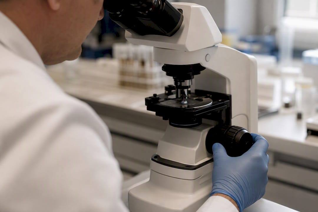
Architectural features associated with melanoma include disarrayed epidermal architecture that breaks the normal honeycomb pattern, non-edged papillae (dermal papillae that lack the sharp, bright rim seen in benign nevi), atypical melanocytic nesting, and irregular junctional patterns where the dermoepidermal border loses its usual regularity.
Cytologic features carry even more diagnostic weight. Melanoma cells frequently appear pleomorphic and brightly nucleated, standing out sharply from the more uniform cells of benign nevi. Pagetoid spread, meaning atypical roundish cells migrating up into the epidermis outside the basal layer, is one of the more specific signs clinicians learn to spot. Some melanoma subtypes also show abundant bright dendritic cells scattered through the epidermis, a feature that requires careful distinction from normal Langerhans cells.
Distinguishing melanoma from its common mimics is where scoring systems earn their keep, discussed further below. A few patterns are worth knowing in advance:
- Solar lentigo typically shows a regular, uniform honeycomb pattern with edged papillae and lacks the cytologic atypia seen in melanoma.
- Lichen planus-like keratosis can show inflammatory infiltrate and some architectural disruption, which sometimes confuses even experienced readers, but it generally lacks true pagetoid melanocytic spread.
- Seborrheic keratosis presents with characteristic keratin-filled crypts and a cerebriform surface pattern that differs structurally from melanocytic proliferation.
Small-diameter melanomas deserve a specific mention. Lesions under 6mm can be dermoscopically unremarkable yet still show subtle cytologic atypia on RCM, which is one of the technology’s more underappreciated strengths. The same holds for amelanotic lesions, where a scattering of unexpectedly bright cells can be the only clue pointing toward malignancy before histopathology confirms it.
What Are the Limitations of Confocal Microscopy for Melanoma?
RCM is not a staging tool, and treating it like one is the single most consequential misuse of the technology. It cannot reliably measure Breslow thickness or assess deep dermal invasion, because imaging depth tops out around the upper dermis. A lesion that looks confined and reassuring on RCM can still harbor deeper invasion that only histopathology will catch.
Surface conditions are the next major obstacle. Hyperkeratosis, crust, blood, and ulceration scatter and absorb the near-infrared light before it reaches diagnostically useful depth, which routinely produces uninterpretable images on exactly the lesions clinicians most want answers about. Acral skin presents a related problem: thicker stratum corneum on palms and soles limits how deep the signal can penetrate, reducing RCM’s reliability at these sites.
Interpretation carries its own traps, and they are well documented rather than theoretical:
- Dendritic cell misidentification. Langerhans cells and atypical melanocytic dendritic processes can look similar under RCM, and confusing the two skews interpretation in either direction.
- Nevoid-type melanomas. These can appear deceptively bland and low-grade on RCM, mimicking benign nevi closely enough to produce false negatives.
- Nodular and ulcerated melanomas. Both tend to image poorly and are a recognized source of false-negative readings, reinforcing why these presentations should bypass RCM in favor of direct biopsy.
- Operator and experience dependence. Diagnostic accuracy correlates strongly with the reader’s years of RCM experience, meaning the same lesion can be read differently by a novice and an expert.
Pro Tip: If dermoscopy and clinical exam both point toward malignancy but RCM comes back reassuring, trust the clinical picture. Discordant findings should trigger a biopsy, not a sigh of relief. RCM works best as confirmation, not veto power, over strong clinical suspicion.
How Do You Fit RCM Into a Dermatology Clinic Workflow?
Adopting RCM is less about buying equipment and more about redesigning a few minutes of every relevant patient visit.
- Time and throughput. Handheld probes like the VivaScope 3000 typically image a lesion in a few minutes, suited to same-visit decision-making. Static mosaic systems like the VivaScope 1500 take longer per lesion because they capture wide-field maps, but they generate more comprehensive documentation for complex or large lesions.
- Training and credentialing. Meaningful proficiency requires a real case volume, not a weekend course. Mentorship arrangements or teleconsultation review with an experienced RCM reader during the early learning curve measurably improve accuracy and shorten the ramp-up period.
- Cost considerations. Capital cost for static systems is substantial, while handheld units carry a lower upfront cost but still require per-case time investment. The trial evidence on reduced excisions suggests RCM can be cost-effective over time by cutting the volume of benign lesions sent to surgery.
- Integration. RCM images need a home. Practical workflow means storing images alongside dermoscopic photos and eventual pathology reports, ideally in a system that lets a dermatopathologist review all three together rather than in isolation.
The clinics that get the most value tend to treat RCM as one more data stream feeding a single decision, not a standalone verdict machine.
What Scoring Systems Help Interpret Confocal Images?
Several validated algorithms convert RCM’s rich visual information into a reproducible score, which matters enormously for consistency across different readers.
The Pellacani score, sometimes called the Modena algorithm, evaluates a defined set of architectural and cytologic features and assigns points for each. In some cohorts, a threshold score of 3 or higher has been linked to sensitivity around 92% and specificity around 72%, figures that track closely with the pooled meta-analysis numbers discussed earlier. The Barcelona algorithm takes a similar feature-based approach but weights certain criteria differently, and the LM score was developed specifically for lentigo maligna, a lesion category where standard scoring systems often underperform.
The practical implication is straightforward: a score threshold gives clinicians a defensible, reproducible cutoff for biopsy versus monitoring, rather than relying purely on gestalt impression. That consistency matters most for less experienced readers, where a structured score can partially compensate for a shorter learning curve.
None of these algorithms were designed to run in isolation. Every validation study behind them assumed the score would sit alongside clinical history and dermoscopic findings, not replace either one. Treating a Pellacani score of 2 as a green light to skip biopsy on a lesion the clinician still finds worrisome ignores exactly what the evidence supports.
How Should RCM Findings Be Documented and Reviewed?
A defensible RCM workflow needs a report structure and a quality-assurance rhythm, not just a good image.
A useful report template covers clinical context (patient history, lesion duration, relevant risk factors), the dermoscopic impression that prompted imaging, RCM findings broken into architectural and cytologic observations, the algorithm score if one was applied, an overall interpretation, and a specific management recommendation.
Quality assurance holds the whole system together over time:
- Peer review of ambiguous or borderline cases before finalizing a management decision.
- Regular case review meetings where readers compare interpretations against eventual histopathology outcomes.
- Systematic image archiving so past lesions can be revisited if new symptoms emerge.
- Periodic calibration exercises, especially for less experienced readers, to catch drift in interpretation standards.
The record itself should document shared decision-making clearly, particularly when the plan is monitoring rather than immediate biopsy. A one-line note that the patient understood and accepted a surveillance plan, with a defined follow-up interval, protects both the patient and the clinician.
A Practical Checklist for Using Confocal Microscopy on a Suspicious Lesion
A concise sequence keeps the decision process consistent across a busy clinic day.
- Select the patient. Confirm the lesion is genuinely equivocal after dermoscopy, not nodular, ulcerated, or overtly high-suspicion.
- Prepare the lesion. Clean without leaving residual moisture or antiseptic film that scatters the imaging beam.
- Image in sequence. Use a handheld probe for a quick screen or a static mosaic system for a full lesion map, depending on size and location.
- Score the findings. Apply Pellacani, Barcelona, or LM score criteria as appropriate to the lesion type.
- Integrate with dermoscopy. Cross-check RCM findings against the dermoscopic impression that prompted the imaging in the first place.
- Document and decide. Record findings using a structured template and commit to a clear plan.
The decision splits three ways: excise immediately when RCM and clinical suspicion align on malignancy, monitor with digital dermoscopy when both point toward benignity, or schedule short-interval RCM follow-up when the picture stays genuinely mixed. A documentation snippet as simple as “RCM Pellacani score 2, dermoscopy reassuring, plan: digital monitoring at 3 months” takes seconds to write and protects the clinical reasoning behind the decision.
How EIV Diagnostics Supports RCM-Based Melanoma Workups
Confocal imaging findings only carry weight when they connect cleanly to the rest of the diagnostic chain. That is where a dedicated pathology partner earns its keep. EIV Diagnostics offers dermatopathology services built to receive biopsy specimens flagged by RCM findings and return board-certified interpretation without the delays that come from routing samples through a generalist lab.
For lesions where RCM findings are ambiguous or where a second opinion strengthens the record, EIV Diagnostics also provides histopathology and digital pathology services that let referring clinicians share high-resolution slide images for consultative review. When a melanoma workup calls for deeper molecular characterization, molecular pathology support is available to complement the histologic diagnosis.
Practically, this means a clinic running RCM on suspicious lesions has a straightforward referral path: submit the biopsy specimen with clinical and imaging context, and expect a board-certified pathologist’s report that closes the loop between what the confocal images suggested and what the tissue actually shows. That connective tissue between imaging and pathology is often the weakest link in RCM-driven melanoma programs, and it is one EIV Diagnostics is built specifically to reinforce.
Where RCM Fits in the Bigger Picture of Melanoma Care
The gap between what RCM promises and what it actually delivers is smaller than either the skeptics or the enthusiasts suggest. It is not a screening replacement for a thorough skin exam, and it is not the diagnostic endpoint dermoscopy critics sometimes want it to be. It is a genuinely useful filter that catches ambiguity dermoscopy alone cannot resolve.
The technology’s future probably runs through better integration rather than dramatic reinvention. Smaller, more affordable handheld devices and early attempts at pairing RCM images with pattern-recognition software could shorten the learning curve that currently gatekeeps accuracy. None of that changes the core lesson from the trial and meta-analysis data available now: RCM’s most reliable role is as an adjunct that sharpens the biopsy decision, not a tool that replaces clinical judgment or pathology. Clinics that adopt it with real training investment and honest quality assurance get real value. Clinics that treat it as a shortcut around biopsy for worrisome lesions are misusing a genuinely good tool.
Referring Suspicious Lesions for Pathology Confirmation
Confocal findings are only as useful as the pathology report that follows them. When a lesion crosses the threshold into biopsy territory, whether RCM pointed there directly or clinical suspicion overrode a reassuring image, EIV Diagnostics gives referring clinicians a fast, direct path to board-certified interpretation.

EIV Diagnostics runs an independent lab model built around dermatopathology, histopathology, digital pathology, and molecular pathology, so a single melanoma workup does not have to bounce between disconnected vendors. Submission is straightforward: send the specimen with clinical history and RCM or dermoscopic findings attached, and a board-certified pathologist returns a report built to integrate with the imaging context you already have. For clinics that also need patient-facing lab draws without adding front-desk logistics, mobile phlebotomy rounds out the workflow. If a suspicious lesion needs tissue confirmation, start by reviewing dermatopathology services and submit the case directly.
Key Studies and Reviews Worth Reading
Clinicians building an evidence-based RCM protocol should start with the primary sources behind the numbers cited throughout this article, rather than secondhand summaries.
- The JAMA Dermatology randomized clinical trial remains the strongest evidence available, since it is one of the few RCT-level studies in dermatologic imaging and directly measures the excision-rate impact clinicians care about most.
- The Cochrane-style systematic review on diagnosing cutaneous melanoma in adults offers the clearest head-to-head comparison of RCM against dermoscopy alone.
- The Wiley meta-analysis across 32 studies gives the most robust pooled sensitivity and specificity figures, along with the setting-dependent variation every reader should understand before quoting a single number.
- The PMC review of benefits and pitfalls is essential reading for anyone implementing RCM clinically, since it catalogs the false-negative patterns and interpretation traps in plain terms.
- The technical overview of RCM’s clinical applications explains the optical principles and imaging depth in enough detail to ground the accuracy data in physical reality.
This article is general information, not a substitute for advice from a qualified doctor. Consult a qualified healthcare professional about your own circumstances before acting on anything here.
Sources
- Effect of Reflectance Confocal Microscopy for Suspect Lesions on Diagnostic Accuracy in Melanoma: A Randomized Clinical Trial
- Reflectance confocal microscopy diagnostic accuracy for malignant melanoma in different clinical settings: systematic review and meta-analysis
FAQ
What does melanoma look like under a microscope?
On reflectance confocal microscopy, melanoma typically shows disarrayed epidermal architecture, non-edged dermal papillae, pagetoid roundish cells, and pleomorphic, brightly nucleated cells that stand out from the uniform pattern seen in benign nevi.
What is the two-week rule for melanoma?
The two-week rule refers to urgent referral pathways in some healthcare systems that guarantee a specialist dermatology appointment within two weeks for lesions with features highly suspicious for melanoma, prioritizing rapid biopsy over prolonged monitoring.
Can confocal microscopy be used to detect skin cancer?
Yes. Reflectance confocal microscopy is a validated adjunct for detecting melanoma, with pooled sensitivity around 92% and specificity near 70%, and it also helps evaluate other pigmented and non-pigmented skin lesions when paired with clinical and dermoscopic assessment.
What causes 90% of melanoma cases?
Ultraviolet radiation exposure, whether from sun or tanning devices, is the dominant environmental driver behind the large majority of melanoma cases, which is why lesion changes on chronically sun-exposed skin deserve close monitoring and, when appropriate, RCM evaluation.
Does a reassuring RCM result mean a lesion is definitely benign?
No. RCM reduces unnecessary excisions but does not eliminate false negatives, particularly for nodular, ulcerated, or nevoid-type melanomas, so a reassuring RCM finding should never override strong clinical or dermoscopic suspicion.
Recommended
- Confocal Microscopy Services | EIV Diagnostics | EIV Diagnostics | EIV Diagnostics
- Dermatopathology Consultations: A Patient’s Guide | EIV Diagnostics | EIV Diagnostics
- Molecular Diagnostic Testing: What Patients and Clinicians Need to Know | EIV Diagnostics | EIV Diagnostics
- Histopathology Services | EIV Diagnostics | EIV Diagnostics | EIV Diagnostics