
By EIV Diagnostics · October 7, 2026
Reflectance Confocal Microscopy for US Clinics: Evidence, Billing, Training
Implement reflectance confocal microscopy: pooled melanoma accuracy, CPT 96931–96936 billing, practical training steps, and EIV Diagnostics lab support.
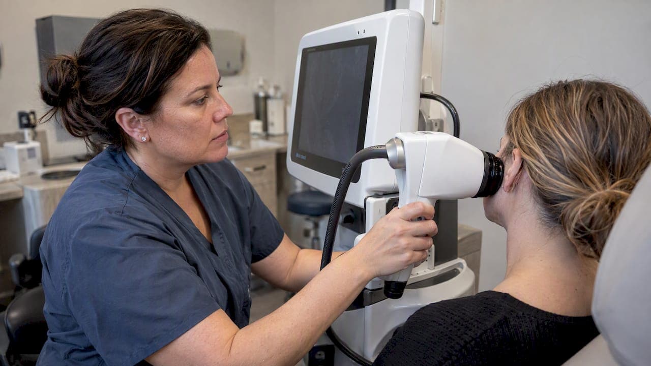
Reflectance confocal microscopy (RCM) is a noninvasive, in vivo, near-histologic imaging technique that meaningfully increases specificity for suspicious skin lesions and helps guide biopsy and surgical planning. Pooled data put melanoma sensitivity near 94% and specificity near 76%, and CPT codes 96931 through 96936 now cover its use in U.S. practice. Consider RCM for equivocal pigmented lesions or margin mapping before deciding whether to biopsy.
TL;DR:
- RCM offers high sensitivity for melanoma detection at around 94%, but its specificity of about 76% means it reduces unnecessary biopsies by ruling out benign lesions.
- Its imaging depth of approximately 200 micrometers limits assessment to superficial and junctional skin processes, making it unsuitable for deeply invasive or nodular tumors.
- Operator experience and image quality significantly influence diagnostic accuracy, while high equipment costs and limited availability hinder widespread adoption.
- RCM is best used as a triage tool to clarify ambiguous pigmented lesions, map lesion margins, or guide biopsy site selection, rather than a sole diagnostic method.
- Billing for RCM involves CPT codes 96931-96936, with reimbursement varying locally, and effective implementation depends on proper staff training and integration with pathology services.
Table of Contents
- What RCM Images and What It Leaves Out
- How RCM Works: Optics, Contrast, and Reading the Images
- Where RCM Changes Clinical Decisions
- What the Pooled Evidence Actually Shows
- Where RCM Falls Short
- Capturing, Documenting, and Billing an RCM Exam
- Building RCM Into Your Practice
- How EIV Diagnostics Supports RCM Workflows
- Safety and Who Should Avoid RCM
- Reading RCM Images Consistently
- Where the Technology Is Headed
- Clinician Perspective: Adoption, Barriers, and What’s Next
- Getting Started With Confocal Microscopy Through EIV Diagnostics
- FAQ
- Sources
What RCM Images and What It Leaves Out
Reflectance confocal microscopy is an in vivo, cellular-resolution imaging method that produces en face (horizontal) views of skin, roughly parallel to the surface rather than the vertical cross-sections a pathologist sees under a microscope. It uses a low-power near-infrared laser to generate grayscale images at a resolution fine enough to show individual keratinocytes, melanocytes, and nuclear outlines, which is why clinicians often call it an optical or “light” biopsy. According to a clinical review of RCM principles and uses, the technique is best understood as an adjunct to clinical and dermoscopic assessment rather than a stand-alone diagnostic tool.
The imaging depth is the key constraint to keep in mind. RCM reaches approximately 200 micrometers, which covers the epidermis down to the papillary dermis but stops well short of the reticular dermis or subcutaneous fat. That means superficial and junctional processes are well captured, while deeply invasive or nodular lesions, and anything below the papillary dermis, fall outside its reach. A clinician evaluating a lesion that seems to extend into deeper tissue on physical exam should not expect RCM to characterize that depth.

Commercial RCM systems are built around point-scanning confocal optics, with handheld and tabletop configurations available from a small number of manufacturers; VivaScope-branded devices are the most commonly referenced in published literature and clinical practice, though device selection itself is a separate decision from the clinical question RCM is meant to answer. For a shorter introduction to how this fits into routine skin diagnosis, see our overview of confocal microscopy for skin diagnosis.
The practical takeaway: RCM gives you a magnified, near-histologic look at the epidermis and upper dermis without cutting the skin, and it works best as a triage step before biopsy, not as a replacement for histopathology in cases where deeper assessment matters.
How RCM Works: Optics, Contrast, and Reading the Images
RCM optics rely on a confocal principle: a near-infrared laser, typically around 830 nanometers, is focused onto a single point in the tissue, and a pinhole aperture in front of the detector rejects light scattered from outside that focal plane. The result is an image built almost entirely from light reflected at one depth, which is what allows RCM to produce sharp, cellular-level detail instead of a blurred composite of multiple tissue layers. The system scans this focal point across the tissue in a raster pattern, assembling thousands of point measurements into a single en face grayscale image.
Contrast in RCM comes from differences in the refractive index of structures within the skin rather than from dyes or stains. Melanin is the strongest natural reflector in skin, so melanocytes, melanin-laden keratinocytes, and pigmented structures appear bright against a darker background. This is precisely why RCM has found its strongest foothold in pigmented lesion assessment. Keratin within the stratum corneum and spinous layer produces a honeycomb or cobblestone pattern that experienced readers use as a landmark for normal epidermal architecture. Collagen in the papillary dermis produces a characteristic bright, fibrillar pattern that helps orient the reader to the dermal-epidermal junction, which is often the single most informative plane in a pigmented lesion workup.
Because RCM images are acquired en face rather than in vertical cross-section, they do not map directly onto the tissue planes a dermatopathologist examines on an H&E slide. A clinician trained on histopathology has to learn a new mental model: instead of looking down through skin layers in a single static image, RCM reading involves scrolling through sequential horizontal planes at increasing depth, roughly analogous to flipping through stacked floor plans of a building rather than viewing its elevation.
Most clinically useful RCM examinations are not single images but mosaics, stitched composites of multiple adjacent fields captured at a given depth to cover an entire lesion rather than a small sample of it. Mosaicking is particularly important for margin mapping, where the goal is to characterize the full peripheral extent of a lesion rather than a central hot spot. Image labeling, consistent orientation, and depth annotation during acquisition matter here because a mosaic that is poorly indexed becomes difficult to correlate later with a biopsy site or a follow-up image.
Where RCM Changes Clinical Decisions
RCM earns its place in a workup when the clinical or dermoscopic picture is ambiguous enough that the next step would otherwise be a biopsy with uncertain yield. Its most established applications include:
- Pigmented lesion triage: RCM adds a cellular-level layer of evidence to dermoscopy for lesions that look atypical but not definitively malignant, helping decide whether to excise or monitor.
- Lentigo maligna margin mapping: mapping the often subclinical, irregular extent of lentigo maligna before Mohs or staged excision, since these lesions frequently extend well beyond their visible pigmented borders.
- Basal cell carcinoma assessment: both initial diagnosis and margin or residual tumor assessment after treatment, particularly for superficial and nodular subtypes within the imaging depth.
- Non-oncologic uses: evaluating inflammatory dermatoses, some infectious lesions, and mapping scalp or widespread pigmented macules to select the most representative biopsy site.
For lentigo maligna specifically, RCM-guided margin mapping shows high agreement with histology when used to delineate subclinical extension, which supports its growing role in presurgical planning for this notoriously ill-defined tumor type. The same logic extends to biopsy-site selection in multifocal presentations: a clinician facing several similar-looking pigmented macules, or a patchy area of scalp hair loss, can use RCM to identify the single area most likely to be diagnostic, reducing the odds of a non-diagnostic or misleading biopsy. Our melanoma-focused review of confocal microscopy’s accuracy, uses, and limits covers the pigmented lesion application in more depth.
In practice, the clinical question that matters most is whether RCM findings would change management, whether that means proceeding to excision, choosing active monitoring, or redirecting a biopsy to a better location.
What the Pooled Evidence Actually Shows
Meta-analytic data put RCM’s pooled sensitivity for melanoma at roughly 94% (95% CI 0.87 to 0.98) and specificity at roughly 76% (95% CI 0.67 to 0.85), according to a head-to-head comparison of RCM and dermoscopy. That specificity figure is the number worth sitting with: it means RCM correctly rules out a meaningful share of lesions that dermoscopy alone would flag as suspicious, translating into fewer biopsies performed on benign lesions without a comparable loss in melanoma detection.

For basal cell carcinoma, a systematic review and meta-analysis of RCM diagnostic accuracy covering 15 studies and 4,163 lesions reported pooled sensitivity of 0.92 (95% CI 0.87 to 0.95) and pooled specificity of 0.93 (95% CI 0.85 to 0.97). Both sensitivity and specificity cluster in the low-to-mid 90s for BCC, which is a stronger combined performance than RCM shows for melanoma, consistent with BCC’s more stereotyped confocal appearance (elongated monomorphic nuclei, increased dermal vascularity, and polarized basaloid structures).
The gap between RCM and dermoscopy largely shows up on the specificity side rather than sensitivity. Dermoscopy alone tends to flag more lesions as atypical than later prove malignant; RCM’s cellular-level view lets clinicians rule out a substantial share of those false alarms before committing to a biopsy. Reviews addressing this comparison have estimated that RCM-guided triage can reduce unnecessary excisions in equivocal lesions by roughly 30 to 40% in selected cohorts, though that range reflects heterogeneous study populations and should be read as a directional finding rather than a fixed rate for any individual practice.
The width of the confidence intervals, particularly for melanoma specificity (0.67 to 0.85), signals real heterogeneity across the underlying studies. Three factors drive most of it: case mix (a dermatology referral center seeing a high proportion of genuinely atypical lesions will show different performance than a general practice screening a broader population), operator experience (readers with more years of RCM interpretation consistently perform better in comparative studies), and the mix of lesion subtypes included (amelanotic or poorly pigmented melanomas are harder to characterize than classic pigmented lesions). None of this undermines the overall direction of the evidence, but it does mean a single practice’s results will not necessarily mirror the pooled averages exactly.
Where RCM Falls Short
RCM’s limitations trace directly back to its physics. The roughly 200-micrometer imaging depth means deeply invasive melanomas, nodular basal cell carcinomas with significant depth, and most non-epidermal processes are poorly visualized or missed entirely; a lesion that needs deeper characterization still needs a biopsy regardless of what RCM shows at the surface.
Operator and reader variability is a second, less obvious limitation. Image acquisition technique, pressure artifact, and motion blur can all distort cellular architecture in ways that mimic atypia, and less experienced readers are more prone to both false positives (over-calling benign lesions as suspicious) and false negatives (missing subtle atypical features). A clinical review of RCM in practice notes that reader experience is itself a measurable source of diagnostic variability, not just a theoretical concern.
Practical constraints matter just as much as technical ones. RCM devices carry significant upfront cost, imaging sessions run longer than a standard dermoscopic exam, especially for large lesions requiring extensive mosaicking, and equipment availability remains limited outside academic and specialty referral centers. These constraints are a major reason RCM has not become a universal screening tool despite strong performance data: the technique is better suited to a focused, equivocal-lesion workflow than to high-volume general use.
Capturing, Documenting, and Billing an RCM Exam
A usable RCM exam depends on acquisition discipline as much as on the equipment itself. A practical sequence looks like this:
- Localize the lesion with standard photography and dermoscopy first, so the RCM field of view can be registered to a known anatomic landmark.
- Capture mosaics at each clinically relevant depth, typically the stratum corneum, spinous-granular layer, and dermal-epidermal junction, rather than isolated single frames.
- Label and archive images with consistent depth and orientation markers so a second reader or a future follow-up visit can locate the same field.
- Document interpretation in a structured note that separates descriptive findings (architecture, cytology, vascular pattern) from the overall impression.
A written RCM report should give a referring clinician enough to act on without re-reading the raw images: a brief description of architectural and cytologic findings at each depth, a stated impression (benign, equivocal, or suspicious), and a recommendation (observe, biopsy, or re-image). When a finding is borderline, requesting a second opinion from another RCM-trained reader, or correlating directly with a pathology partner, is standard practice rather than an exception. Our dermatopathology services page describes how that kind of cross-check with histopathology typically works.
On the billing side, CPT Category I codes 96931 through 96936 cover reflectance confocal microscopy, and they separate into technical and professional components, meaning the image acquisition and the physician interpretation can be billed distinctly depending on who performs each step. According to CMS’s physician fee schedule lookup, Medicare reimbursement for these codes varies by locality and by year’s fee schedule update, so practices need to verify current rates for their specific Medicare Administrative Contractor rather than assume a flat national rate, and private payer coverage should be confirmed separately before scheduling a patient for RCM.
Building RCM Into Your Practice
Adding RCM to a practice is less about the purchase order and more about the training runway. A workable sequence looks like this:
- Start with a structured course. Single-center reports on CME-accredited RCM courses in the United States describe strong gains in interpretive confidence after short, structured training programs, with many attendees going on to integrate RCM into clinical practice afterward.
- Decide on a staffing model. Some practices train a dedicated technician to handle image acquisition while the clinician focuses solely on interpretation; others have the same clinician acquire and read every exam. Either model works, but mixing acquisition quality across untrained staff is a common source of avoidable artifact.
- Build a correlation habit. Every early RCM read should be checked against the eventual histopathology result when a biopsy follows, creating a local feedback loop that sharpens interpretation faster than reading in isolation.
- Maintain a local image library. Archiving representative benign, equivocal, and malignant images from your own practice gives new readers a reference set that matches the patient population you actually see.
Pro Tip: Budget a protected period of several months, not weeks, for new readers to build real concordance with histopathology before relying on RCM impressions to change management independently.
Dedicating staff specifically to acquisition, rather than treating it as an occasional add-on task, measurably reduces the artifact rate that otherwise erodes reader confidence early on.
How EIV Diagnostics Supports RCM Workflows
Our diagnostic services are built around the reality that imaging and pathology need to talk to each other, not sit in separate systems. We offer dermatopathology, digital pathology, and confocal microscopy services alongside mobile phlebotomy for specimen collection, with pathologists reading cases and reporting findings.
For a practice adding RCM, the value of a pathology partner shows up at the correlation step: when an RCM impression needs to be checked against histopathology, or when a referring clinician wants a second opinion on a borderline read, having a lab that handles both dermatopathology and digital pathology under one roof shortens that loop considerably. Cases are reported by pathologists and quality standards are maintained across testing, which matters when RCM findings are being used to decide whether a lesion needs a biopsy at all.
Safety and Who Should Avoid RCM
Reflectance confocal microscopy uses a low-power near-infrared laser and does not involve ionizing radiation, injected contrast, or tissue removal, which is a meaningful part of its appeal for cosmetically sensitive sites like the face. The laser power used in commercial systems is designed to stay below levels associated with thermal tissue damage, and the exam itself is generally well tolerated, with mild pressure from the imaging tip against the skin being the main physical sensation reported.
Open wounds, active infection at the imaging site, or extremely thin or fragile skin can make acquiring a usable image difficult or uncomfortable, and these situations are better handled by waiting for the skin to stabilize or choosing a different diagnostic approach first. Lesions suspected to be deeply invasive on physical exam are a relative contraindication in the sense that RCM will not meaningfully inform management; the exam effort is better spent proceeding directly to biopsy. There is no established need to avoid RCM in pregnancy or in patients with photosensitivity, since the device does not use ultraviolet light, though any device-specific instructions from the manufacturer and standard infection control between patients should be followed as a matter of routine clinical practice.
Reading RCM Images Consistently
Because RCM produces unfamiliar en face views rather than traditional histologic cross-sections, consistent interpretation depends on checking the same structural features at each depth rather than forming an overall gestalt impression from a single frame. A structured read typically moves from the stratum corneum downward, noting keratinocyte and nuclear morphology, the regularity of the honeycomb pattern at the spinous-granular layer, and then the architecture at the dermal-epidermal junction, where pigmented lesion atypia is most often visible.
At the junction, readers specifically look for ringed versus non-edged dermal papillae, the regularity of junctional melanocytic nests, and the presence of pagetoid cells (atypical melanocytes scattered above the basal layer), since these features carry the most weight in distinguishing benign nevi from melanoma on RCM. For BCC, the pattern shifts toward elongated monomorphic basaloid nuclei, polarization along a common axis, and increased dermal vascularity.
A written impression should state findings separately from the overall assessment rather than blending description and conclusion into one sentence, which makes it easier for a second reader or a referring clinician to independently evaluate the same images later. Reading in isolation from the clinical and dermoscopic context is a common mistake: RCM findings are meant to be interpreted alongside the lesion’s history and dermoscopic appearance, not as a stand-alone verdict.
Where the Technology Is Headed
The most active area of RCM development is artificial intelligence-assisted image analysis, aimed at flagging suspicious regions within a mosaic and reducing the reader-experience gap that currently drives much of the variability in diagnostic accuracy. Automated feature detection, trained to recognize the same architectural patterns experienced readers look for, is being explored as a way to standardize first-pass reads before a human confirms the impression.
Device miniaturization is the second major trend, with handheld units aimed at making RCM practical outside dedicated imaging suites and more accessible for bedside or satellite-clinic use. Combined reflectance and fluorescence confocal systems are also in development, intended to add a second contrast mechanism on top of the native reflectance signal for better discrimination of certain cell types.
Teleconsultation and remote image review are gaining traction as a way to extend RCM access beyond the academic centers that currently have the equipment and trained readers, letting a smaller practice acquire images locally while a remote specialist interprets them, a pattern already well established in remote-reading workflows for other imaging modalities. None of these developments change RCM’s fundamental depth limitation, but together they target the two biggest adoption barriers: reader variability and equipment access.
Clinician Perspective: Adoption, Barriers, and What’s Next
The honest case for RCM adoption is narrower than the marketing around it suggests. The technology earns its place for one specific reason: it moves the needle on specificity for equivocal pigmented lesions, which translates into real, measurable reductions in unnecessary excisions. That is a genuine clinical win, not a convenience feature.
The barriers holding back wider adoption are mundane rather than scientific: training time, equipment cost, and reimbursement uncertainty that varies by payer and locality. Those barriers will erode faster than the core technology improves. AI-assisted reading, handheld devices, and teleconsultation models are all aimed at the access and variability problems, not at some fundamental flaw in RCM itself. Clinicians waiting for a dramatic technical leap before adopting RCM are, in our view, waiting for the wrong thing: the evidence already supports selective use today.
— EIV Diagnostics
Getting Started With Confocal Microscopy Through EIV Diagnostics
We make it straightforward for clinicians to add confocal microscopy to an existing workup without building an entire imaging program from scratch. Whether you need imaging support for an equivocal pigmented lesion, margin mapping before a planned excision, or a second opinion that ties an RCM impression back to histopathology, our lab is set up to handle that handoff directly.

Clinicians and practices can reach us through the following services:
- Confocal Microscopy Services: order confocal imaging as a standalone service or alongside a pathology referral.
- Dermatopathology Services: correlate RCM findings with histopathology read by board-certified pathologists.
- Digital Pathology: share and review case images remotely as part of a second-opinion workflow.
- Mobile Phlebotomy: arrange specimen collection at a patient’s home or office when a biopsy follows an RCM exam, from $65 one-off.
Reach out to our team to discuss integrating confocal microscopy and dermatopathology reporting into your practice’s workflow.
This article is general information, not a substitute for advice from a qualified doctor. Consult a qualified healthcare professional about your own circumstances before acting on anything here.
FAQ
Can a dermatoscope see cancer?
A dermatoscope magnifies surface and subsurface pigment patterns but cannot show cellular-level detail the way reflectance confocal microscopy or histopathology can. It raises or lowers suspicion for cancer based on pattern recognition, which is why equivocal dermoscopic findings are often followed by RCM or biopsy rather than a dermatoscope-only diagnosis.
What is the purpose of confocal microscopy?
Reflectance confocal microscopy provides noninvasive, cellular-resolution imaging of the epidermis and upper dermis, functioning as an optical “light biopsy” for equivocal skin lesions. Its main purpose is to increase diagnostic specificity before deciding whether a lesion needs excision, as described in a clinical review of RCM in practice.
What are the downsides of using a confocal microscope?
RCM’s imaging depth of roughly 200 micrometers means it cannot reliably characterize deeply invasive or nodular lesions, and interpretation accuracy depends heavily on operator and reader experience. Equipment cost, longer appointment times, and limited availability outside specialty centers are the main practical constraints.
What are the different types of confocal microscopy?
Reflectance confocal microscopy, which uses tissue’s natural refractive index differences for contrast, is the form used clinically for in vivo skin imaging. Fluorescence confocal microscopy, which relies on fluorescent dyes or tags, is used mainly in research and some ex vivo pathology applications rather than routine in vivo clinical exams.
Does insurance cover reflectance confocal microscopy?
Coverage depends on the payer and the specific CPT code billed, since CPT codes 96931 through 96936 separate technical and professional components with reimbursement that varies by Medicare locality and private payer policy. Practices should verify coverage with the patient’s specific plan before scheduling an RCM exam.
Sources
- Reflectance confocal microscopy versus dermoscopy for the diagnosis of cutaneous melanoma: a head-to-head comparative meta-analysis
- Introduction to reflectance confocal microscopy and its use in clinical practice
- CMS physician fee schedule search results for CPT 96931-96936