
By EIV Diagnostics · September 2, 2026
Telepathology for Clinics: 7 Step Plan and 60 Case CAP Validation
Practical US guide to telepathology for clinics: 7 step rollout, CAP 60 case validation, costs, EHR integration, and an EIV Diagnostics example.
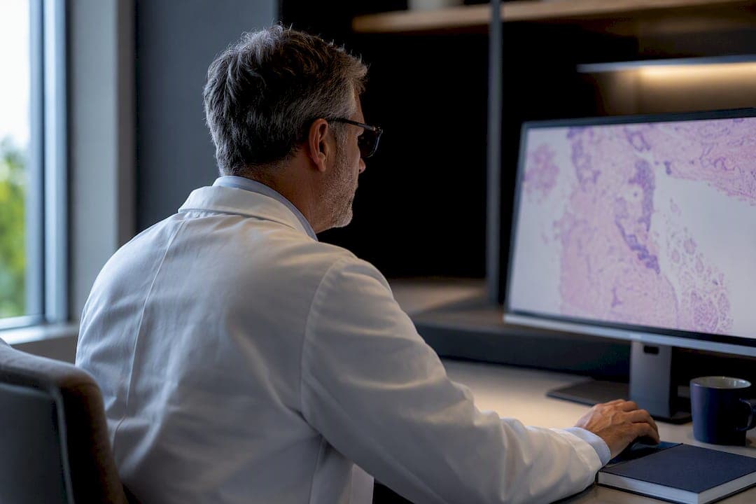
Telepathology lets clinics send pathology images to remote specialists for diagnosis, second opinions, or intraoperative consults without shipping physical slides. It works, and it holds up clinically, but only when a lab validates its whole-slide imaging (WSI) system under College of American Pathologists (CAP) protocols, meets Clinical Laboratory Improvement Amendments (CLIA) obligations, and rebuilds its pre-analytic and post-analytic workflows around digital handoffs. Done right, clinics get faster subspecialty access and shorter turnaround times. Done as a hardware purchase alone, it usually stalls.
TL;DR:
- Validating a whole-slide imaging system under CAP protocols and meeting CLIA requirements is essential before relying on telepathology for primary diagnosis.
- Scanner throughput capacity should match actual case volume, with low-, mid-, and high-throughput options suited for different clinic sizes and needs.
- Integration with existing electronic health records often requires deliberate planning and testing to prevent workflow disruptions and data mismatches.
- Telepathology workflows significantly reduce turnaround times by 30 to 67 percent and maintain diagnostic accuracy above 98 percent compared to traditional microscopy.
- Clinics frequently find partnering with established labs simplifies validation, reduces costs, and accelerates adoption, especially for smaller practices.
Table of Contents
- What Is Telepathology and Which Modalities Do Clinics Use?
- WSI File Sizes, Scanners, and Storage: The Technical Trade-offs
- What Regulatory Requirements Apply to Telepathology in Clinics?
- How Do Clinics Actually Use Telepathology Day to Day?
- A Step-by-Step Implementation Checklist for Clinics
- What Does Telepathology Cost, and How Do You Justify It?
- Managing Risk, Quality, and Cybersecurity in a Telepathology Program
- EIV Diagnostics: A Working Example of Telepathology Integration
- What Happens When Telepathology Meets Your EHR System?
- What Training Do Pathologists and Staff Need?
- Publisher Perspective: Build In-House or Partner With a Lab?
- How EIV Diagnostics Supports Clinics Adopting Telepathology
- Sources
- FAQ
What Is Telepathology and Which Modalities Do Clinics Use?
Telepathology is the practice of transmitting pathology images, either static, streamed, or fully digitized, to a pathologist at another location for diagnosis or consultation. It replaces or supplements the courier-driven model of shipping glass slides between facilities, which can add days to a diagnosis when a subspecialist isn’t on-site.
Three modalities dominate clinical use, and each fits a different situation:
- Static image telepathology. A technician or pathologist captures still images of selected fields under a microscope and sends them for review. It’s cheap and fast to set up but limited by the operator’s choice of fields, so it works best for straightforward consults, not complex primary diagnosis.
- Streaming/robotic microscopy. A remote pathologist controls a motorized microscope in real time over a video connection, moving the stage and adjusting focus as if standing at the scope. This is the workhorse for frozen sections and intraoperative consults, where a live back-and-forth with the surgeon matters.
- Whole-slide imaging (WSI). A scanner digitizes the entire slide into a single high-resolution file that a pathologist reviews on a monitor, at any time, from anywhere. WSI is the modality behind full digital sign-out, tumor board review, and archival case sharing.
Each modality touches a common set of components: a scanner or capture device, an image management system (IMS) to store and route files, integration with the laboratory information system (LIS), a diagnostic-grade workstation, and enough network bandwidth to move large image files without lag. A clinic doing occasional consults might only need static imaging and a secure upload portal. A clinic aiming for full digital primary diagnosis needs a validated WSI pipeline end to end.
WSI File Sizes, Scanners, and Storage: The Technical Trade-offs
The technology decisions here drive both your budget and your day-to-day throughput, so it’s worth understanding what you’re actually buying before a vendor walks you through a demo.
WSI files run large. Depending on scan resolution, a single slide typically produces a file in the 0.5 to 2 GB range, and a busy histology lab can generate hundreds of these files a day. Most clinical scanning happens at 20x magnification, which balances detail against file size and scan speed; some dermatopathology and hematopathology cases call for 40x for finer cellular detail, at the cost of larger files and slower scans. A single slide scan can take anywhere from 60 seconds to several minutes depending on tissue size, magnification, and scanner throughput mode.
Scanner selection should track your case volume, not the flashiest spec sheet:
- Low-volume scanners (single-slide or small-batch loaders) suit clinics doing occasional consults or a handful of frozen sections a week.
- Mid-throughput scanners with 100 to 300 slide capacity fit clinics building toward routine digital sign-out.
- High-throughput scanners with rack-fed automation are built for reference labs and large hospital systems, not typical clinics.
Image management is where interoperability either gets solved or gets ignored until it’s expensive to fix. Some IMS platforms lock files into a proprietary format; others support DICOM for pathology or feed into a vendor-neutral archive (VNA), which keeps your images portable if you ever switch scanner vendors or add a second site. Tiered storage, hot storage for recent active cases and cheaper cold storage for archives, is the standard way labs handle network and storage bottlenecks at scale.
Workstations matter more than clinics expect. A pathologist reading WSI for primary diagnosis needs a calibrated, high-resolution monitor and enough local processing power to pan and zoom a multi-gigabyte file without stutter. Remote access adds another variable: connection latency. The American Telemedicine Association specifically flags diagnostic-grade imaging and low-latency connectivity as prerequisites for clinical reliability, not nice-to-haves.
Pro Tip: Before buying a scanner, run a throughput test with your actual case mix, not the vendor’s demo slides. Mixed tissue types and cellularity affect scan time far more than spec sheets suggest.
What Regulatory Requirements Apply to Telepathology in Clinics?
Regulation here splits along a line that trips up a lot of clinics: device clearance is not the same thing as laboratory validation, and clearing one hurdle doesn’t clear the other.
The FDA classifies WSI systems used for primary diagnosis as Class II medical devices, meaning the scanner and software have cleared a federal review for that intended use. Streaming video systems used for telecytology or informal consults generally fall outside FDA device regulation. But FDA clearance of the hardware doesn’t mean your lab is cleared to use it. Every laboratory still has to independently validate the system in its own hands, on its own case mix, before relying on it for primary diagnosis.
That validation follows CAP guidance, which sets specific thresholds:
- A minimum of 60 cases per preparation type for a lab’s initial WSI validation.
- An additional 20 cases for each subsequent application (a new tissue type, a new stain, a new scanner model).
- Revalidation triggers whenever there’s a major software update, a new scanner, or a significant workflow change.
CLIA obligations sit on top of this and apply regardless of modality. Every clinical laboratory offering digital pathology services carries the same CLIA quality accountability it would for any other testing method: documented quality control, competency assessment, and corrective action procedures. Clinics unfamiliar with the practical side of these requirements can review a primer on CLIA certification for a plain-language breakdown of what’s actually required.
Privacy adds a fourth layer. Image transfer and cloud storage need encryption in transit and at rest, access logging, and vendor HIPAA attestations, since a WSI file is protected health information the moment it’s linked to a patient record.
How Do Clinics Actually Use Telepathology Day to Day?
The workflow looks different depending on what you’re trying to accomplish, and conflating these use cases is a common planning mistake.
- Primary diagnosis sign-out. Full WSI review as the basis for the final diagnostic report. This is the highest-bar use case, requiring completed CAP validation, and most smaller labs approach it gradually rather than all at once; many start with consultative workflows before scaling to primary diagnosis.
- Frozen section and intraoperative consults. Streaming or robotic microscopy connects a remote pathologist to the operating room in real time. Realistic turnaround expectations run 15 to 20 minutes from slide preparation to verbal diagnosis, similar to on-site frozen section timing, provided the network connection holds up.
- Second-opinion and subspecialty consults. A generalist pathologist sends a difficult case to a subspecialist, often across state lines, without shipping the block or slide. This is the lowest-friction entry point for most clinics and the one that delivers value fastest.
- Tumor board integration. Digitized cases get pulled up live during multidisciplinary review, letting surgeons, oncologists, and pathologists look at the same image simultaneously instead of describing findings verbally.
- Quality assurance and education. The same WSI archive used for diagnosis doubles as a teaching and QA resource, letting a lab pull prior cases for peer review or resident training without re-cutting slides.
Case reports and system evaluations show turnaround-time reductions in the range of 30 to 67 percent once telepathology workflows are running, with diagnostic concordance holding above 98 percent against traditional light microscopy in those same reports. That gap between glass and digital is small enough that the operational win, speed, usually outweighs any residual diagnostic variance.
A Step-by-Step Implementation Checklist for Clinics
Most failed telepathology rollouts aren’t technology failures. A lifecycle implementation framework built from real deployments found that workflow redesign and governance, not scanner selection, are the most common points where programs stall. Here’s the sequence that avoids that trap.
- Define governance and scope first. Name a project owner (often a pathologist and an IT lead jointly), set measurable goals (target turnaround time, case types in scope), and decide who signs off on quality issues before a single scanner gets ordered.
- Size the technical build to your actual volume. Match scanner throughput to daily case counts, map out IMS-to-LIS interface requirements, and calculate bandwidth needs for your busiest scanning hours, not your average day.
- Choose vendor-specific or vendor-neutral architecture deliberately. This decision, made early, determines how much flexibility you’ll have later if you add AI tools or a second scanner brand. A vendor-neutral archive costs more upfront and usually pays for itself in avoided lock-in.
- Run the CAP validation study. Cover the required 60-case minimum per preparation type, include a washout period between glass and digital review to avoid recall bias, and document concordance rates formally.
- Train and assess competency before go-live. Pathologists and technicians both need documented sign-off, not just a vendor walkthrough.
- Launch in phases with a glass fallback. Start with one case type or one referring site, keep a parallel glass-slide workflow available during the transition, and set clear service-level agreements for turnaround expectations.
- Track operational metrics and revisit quarterly. Turnaround time, scan failure rate, and case volume per pathologist all tell you whether the system is actually working, not just running.
Pro Tip: Assign a single “digital pathology owner” who is accountable for both the IMPLIES-LIS interface and the validation study. Splitting that ownership between IT and pathology is exactly where the lifecycle framework research found programs lose momentum.
What Does Telepathology Cost, and How Do You Justify It?
Capital costs concentrate in three line items: scanners (ranging widely by throughput tier), IMS software licensing, and diagnostic workstations. Integration work, wiring the IMS into your existing LIS, adds a variable but real cost that clinics frequently underestimate during budgeting.
Operating costs run ongoing and easy to overlook:
- Cloud or on-premise storage, scaled for a growing archive of multi-gigabyte files.
- Network bandwidth sized for peak scanning periods, not averages.
- Vendor service contracts for scanner maintenance and software updates.
- Protected pathologist review time, which the Digital Pathology Association identifies as the most commonly underfunded line item; contractual compensation for remote review time matters as much as any hardware purchase.
On the benefit side, quantify turnaround-time reduction, avoided courier and slide-shipping costs, fewer patient transfers for subspecialty opinions, and any new consult revenue from offering remote second-opinion services. Clinics with tight capital budgets increasingly use subscription or per-case licensing models instead of large upfront scanner purchases, which lowers the barrier to a pilot program significantly.
Managing Risk, Quality, and Cybersecurity in a Telepathology Program
Reliability comes from routine, boring quality control, not from the scanner’s spec sheet.
- Daily and periodic QC. Color calibration checks, slide-level quality checks for scan artifacts (out-of-focus regions, stitching errors), and scheduled scanner maintenance all need documented logs.
- Encryption everywhere. Images need encryption both in transit and at rest, matching the ATA’s guidance on secure transfer as a baseline for clinical reliability.
- Role-based access control. Limit who can view, download, or forward patient images, and require vendor security attestations before onboarding any cloud IMS platform.
- Interoperability safeguards. A vendor-neutral archive and a well-built IMS-LIS interface reduce the risk of getting stuck with unreadable files if a vendor relationship ends.
- Clear accountability across sites. When a remote subspecialist renders a diagnosis for a local patient, the reporting agreement needs to specify who signs the final report and how discrepancies get resolved.
Audit logs tie all of this together: every image access, every diagnostic sign-off, and every QC exception should leave a traceable record.
EIV Diagnostics: A Working Example of Telepathology Integration
EIV Diagnostics, an independent pathology laboratory based in Fresno, California, runs digital pathology services alongside molecular pathology, dermatopathology, and confocal microscopy, giving referring clinics a single point of contact for both digital image review and ancillary testing.
For a clinic integrating with an outside lab, the operational flow generally looks like this:
- Samples get routed either through existing courier arrangements or, for clinics needing draw support, through EIV Diagnostics’ mobile phlebotomy service, which brings specimen collection to the patient’s location.
- Digitized slides and case data move through a validated IMS pipeline to board-certified pathologists for diagnosis.
- Reports return in standard formats designed to integrate with a referring clinic’s existing LIS or EHR workflow, with rapid turnaround built into the reporting agreement.
Case counts matter more than marketing claims here. CAP’s validation standard requires a minimum of 60 cases per preparation type before a lab can rely on WSI for primary diagnosis, the same threshold any credible partner lab should be able to document for its own systems.
For clinics needing subspecialty support beyond general pathology, such as molecular testing tied to a digitized case, that combination of digital review and ancillary diagnostics under one lab relationship cuts down on the coordination overhead of managing multiple outside vendors.
What Happens When Telepathology Meets Your EHR System?
Integration friction shows up almost immediately once a clinic tries to connect a telepathology workflow to its existing electronic health record system. The core problem is architectural: most EHR platforms were built around structured text and coded lab values, not multi-gigabyte image files or the accession-level metadata pathology systems depend on.
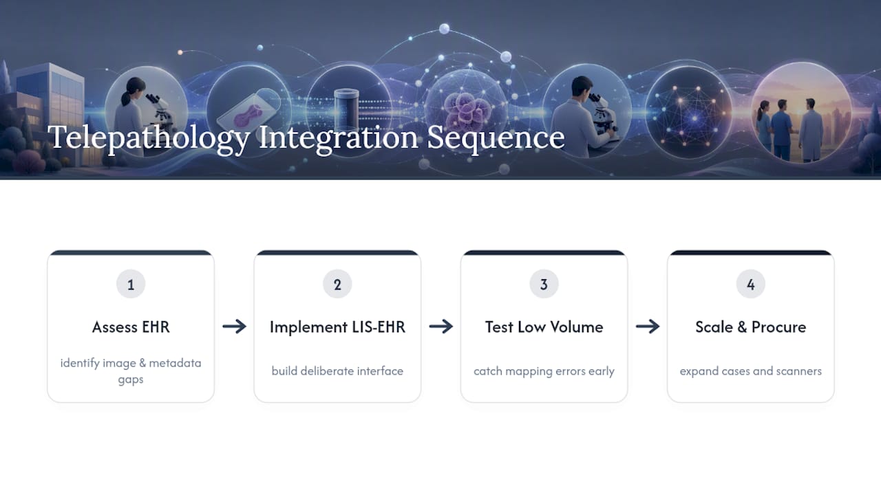
The LIS typically sits between the EHR and the IMS, translating orders and results back and forth, but that interface has to be built and maintained deliberately. A poorly mapped interface can create duplicate patient records, mismatched accession numbers, or delayed result posting, any of which undermines trust in the whole digital workflow faster than a slow scanner would.
Clinics running a smaller EHR platform, common among independent clinics compared to hospital systems, often lack a built-in pathology module at all, which means image links or report PDFs get bolted on rather than natively integrated. That’s a workable interim solution but not a long-term one, since it creates a manual step where staff have to cross-reference systems instead of viewing everything from one chart.
The practical fix is sequencing: build and test the LIS-to-EHR interface before scaling case volume, not after. A lifecycle approach that treats interface work as a distinct project phase, separate from scanner procurement, catches these mismatches while volume is still low enough to fix them cheaply.
What Training Do Pathologists and Staff Need?
Competency for telepathology isn’t the same skill set as reading glass slides under a microscope, and treating it as an afterthought is one of the more common adoption mistakes.
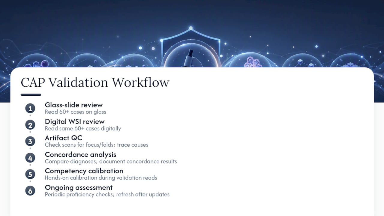
Pathologists reviewing WSI need documented training on navigating digital slides (panning, zooming, focus stacking on multi-layer scans) and on recognizing scan artifacts, like out-of-focus regions or tissue folds, that can mimic or mask real pathology. That training typically pairs with the CAP validation study itself: reading the same 60-plus case set on both glass and digital gives pathologists hands-on calibration time while generating the concordance data the validation requires.
Technical staff need a parallel track. Histotechnologists and scanning technicians need documented competency in slide preparation quality (since scan artifacts often trace back to sectioning or staining issues), scanner operation, and basic troubleshooting for failed or low-quality scans. IT staff supporting the IMS-LIS interface need enough pathology workflow literacy to triage problems without escalating every issue to the vendor.
Ongoing competency assessment matters as much as initial training. CLIA-driven quality programs expect periodic proficiency checks, and any significant software update or new scanner model should trigger a refresher, not just a memo.
Publisher Perspective: Build In-House or Partner With a Lab?
The honest answer depends less on technology comfort and more on volume and capital. High-volume systems planning AI-assisted screening down the road generally justify an in-house build, since they’ll amortize the cost and want architectural control from day one. Smaller and mid-size clinics usually get more value, faster, by partnering with an established lab that has already cleared CAP validation.
A hybrid approach, piloting one use case like second-opinion consults before committing to full digital sign-out, tends to outperform an all-at-once rollout in both directions. Whichever path you choose, insist on a documented validation plan and clear governance before scaling volume.
— EIV Diagnostics
How EIV Diagnostics Supports Clinics Adopting Telepathology
If your clinic is weighing an in-house build against a partner relationship, EIV Diagnostics offers a third path that skips the capital outlay entirely: a working digital pathology pipeline, already validated, staffed by board-certified pathologists, with mobile phlebotomy available for clinics that need specimen collection handled off-site.

Working with EIV Diagnostics means your clinic gets rapid turnaround on digitized cases without buying a scanner, building an IMS from scratch, or running your own CAP validation study. Reports arrive in formats built to slot into your existing referral workflow, and the lab’s molecular pathology and dermatopathology services mean subspecialty testing doesn’t require a second vendor relationship. If you’re evaluating whether to build in-house or partner, start by requesting a service overview through the digital pathology services page and ask specifically about validation support and turnaround guarantees for your case mix.
This article is general information, not a substitute for advice from a qualified doctor. Consult a qualified healthcare professional about your own circumstances before acting on anything here.
FAQ
Is AI Going to Replace Pathologists?
No. AI tools are increasingly used to flag regions of interest or pre-screen images, but diagnostic sign-off still requires a licensed pathologist, and CAP and CLIA frameworks hold the pathologist accountable for the final report regardless of AI assistance.
What Are the Four Types of Pathology?
The four main branches are anatomic pathology (tissue and organ diagnosis), clinical pathology (lab testing of blood and body fluids), molecular pathology (genetic and biomarker testing), and forensic pathology (cause-of-death investigation); EIV Diagnostics offers services spanning several of these, including molecular and dermatopathology.
What Is Telepathology?
Telepathology is the transmission of pathology images, through static capture, live-streaming, or whole-slide imaging, to a pathologist at another location for diagnosis, consultation, or quality review.
How Does Telepathology Work in a Clinical Setting?
A clinic scans or captures slide images, sends them through a secure image management system to a remote pathologist, and receives a diagnostic report back, with workflow speed depending on the modality used and the strength of the underlying network connection.
Who Are Some Vendors for Digital Pathology Products?
Digital pathology vendors range from scanner manufacturers to image management software providers; clinics evaluating vendors should confirm each system’s FDA classification status and request documentation of CAP-compliant validation support before committing to a purchase.