
By EIV Diagnostics · October 4, 2026
Avoid Repeat Biopsies: Skin Biopsy Types and DIF Tips for Clinicians
For clinicians: clear rules for skin biopsy types and practical DIF specimen handling, including Michel's medium and a 3–4 mm punch to improve diagnostic...
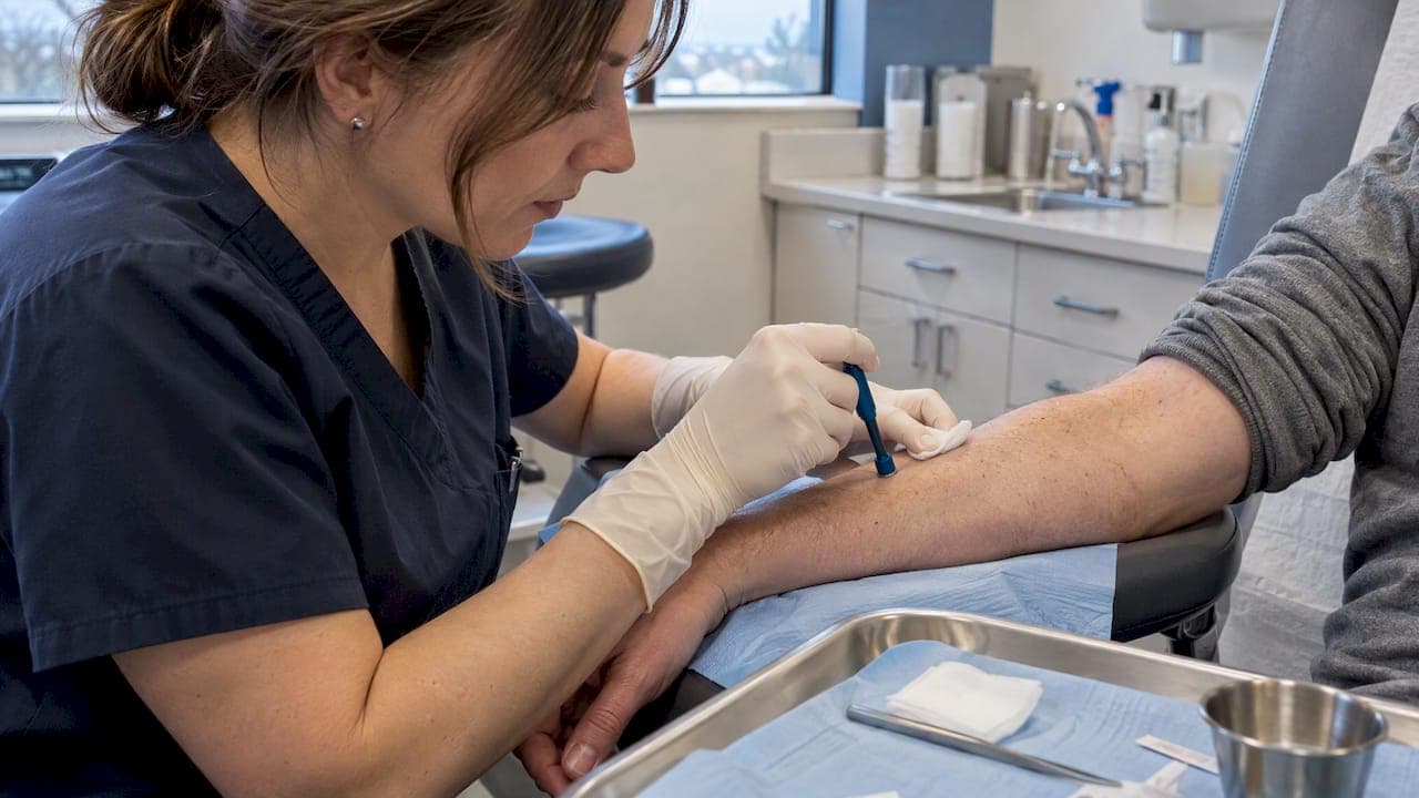
Shave, punch, and excisional or incisional biopsies cover most skin sampling needs, with curette, snip, and saucerization techniques filling specific niches. The right choice depends on how deep the lesion sits, what question the biopsy needs to answer, and whether special testing is involved. Proper handling matters most when additional tests like direct immunofluorescence are needed, which is why labs such as EIV Diagnostics build dermatopathology services around precise specimen protocols.
TL;DR:
- Shave biopsies are ideal for superficial, raised lesions like basal cell carcinomas, but are unsuitable for suspected melanoma due to limited depth assessment.
- Punch biopsies provide full-thickness skin samples suitable for inflammatory conditions, requiring sutures and deeper tissue analysis.
- Excisional biopsies remove entire lesions for comprehensive diagnosis, especially critical when melanoma or border assessment is necessary.
- Special handling is essential for direct immunofluorescence testing, requiring separate samples preserved in Michel’s medium and taken from perilesional, early-stage skin.
- Biopsy choice depends on lesion location, patient skin type, suspicion level, and balancing diagnostic needs with cosmetic and functional outcomes.
Table of Contents
- Overview of the main skin biopsy types
- How clinicians decide which biopsy to use
- What happens during a biopsy and how to care for the site afterward
- Direct immunofluorescence and specialized specimen handling
- Risks, complications, and what healing looks like
- Interpreting biopsy results: what the report means and when to expect it
- EIV Diagnostics: laboratory capabilities built around biopsy accuracy
- Contraindications and limitations for each biopsy type
- Types of anesthesia used during skin biopsies
- Potential complications specific to each biopsy type
- Special considerations for biopsies in different skin types and locations
- Why clinical judgment still drives the choice of biopsy
- Where to get biopsy and specimen support
- FAQ
- Sources
Overview of the main skin biopsy types
Every biopsy technique removes a different slice of skin, and that slice determines what a pathologist can and cannot see under the microscope. Matching the technique to the clinical question is the foundation of an accurate diagnosis, a point reinforced by clinical technique reviews that catalog punch, shave, saucerization, wedge, incisional, and excisional methods as the standard toolkit.
A shave biopsy removes the epidermis and a portion of the superficial dermis using a blade or razor, without cutting into deeper fat or connective tissue. It works well for raised, superficial lesions such as suspected basal cell carcinomas, seborrheic keratoses, or compound moles that sit close to the skin’s surface. Bleeding is controlled with pressure or a topical agent, and stitches are rarely needed, which makes it one of the fastest options in a clinic visit.
A punch biopsy uses a small, tube-shaped cutting instrument, typically 2 to 6 millimeters across, to remove a cylindrical core that reaches full skin thickness, including the dermis and sometimes the upper fat layer. This depth makes it the preferred method for inflammatory skin diseases, unexplained rashes, and conditions where the diagnostic clues sit below the surface. Because the defect goes deeper, a punch biopsy often requires one or two sutures to close.
Excisional and incisional biopsies differ mainly in scope. An excisional biopsy removes an entire lesion along with a margin of normal-looking skin, the standard approach when melanoma is suspected because it lets a pathologist assess the full depth and borders at once, as outlined in MedlinePlus guidance on skin biopsies. An incisional biopsy removes only a representative portion of a larger lesion, useful when complete removal isn’t practical before a diagnosis is confirmed, such as with a large plaque that needs sampling before planning definitive treatment.
Curette, snip, and saucerization techniques round out the list for specific situations.
- Curettage scrapes off superficial growths and works well for some low-risk, well-defined lesions, though it can distort tissue architecture.
- Snip excision uses sharp scissors to clip off pedunculated lesions, such as skin tags, quickly and with minimal anesthesia.
- Saucerization is a deeper, broader shave that captures more of the dermis, often chosen when a standard shave would risk an incomplete margin on a pigmented lesion.
Nail and mucosal biopsies adapt these same principles to tissue that behaves differently. Nail bed or matrix biopsies often use a punch or longitudinal excision and carry a higher risk of permanent nail changes, so they’re reserved for lesions with a real diagnostic need, such as a suspicious pigmented streak. Mucosal biopsies inside the mouth or on genital skin use smaller punches or shave techniques adjusted for thinner, more vascular tissue, with extra attention to hemostasis.
How clinicians decide which biopsy to use
Choosing a technique comes down to a short list of practical questions. Clinicians weigh the lesion’s size and depth, its location on the body, cosmetic concerns, and how strongly they suspect a serious diagnosis like melanoma. A lesion on the face calls for a different trade-off between diagnostic completeness and scarring than one on the back.
- Suspected melanoma: an excisional biopsy with narrow margins is typically chosen so the pathologist can evaluate the entire lesion’s depth and architecture at once.
- Widespread inflammatory rash: a punch biopsy of a representative, well-developed lesion usually gives the clearest picture, since inflammatory patterns depend on seeing the full thickness of skin.
- Large or hard-to-remove lesion: an incisional biopsy samples a piece first, deferring full removal until the diagnosis is clear.
- Superficial, low-suspicion growth: a shave or saucerization biopsy is often enough and avoids unnecessary scarring.
Some presentations call for more than one sample. A patient with blistering skin disease might need both a standard biopsy for routine staining and a second sample handled differently for direct immunofluorescence testing, covered in detail further down. When a clinician anticipates that need, the biopsy plan changes before the first cut is made: site selection, specimen size, and transport medium all depend on knowing in advance which tests the tissue will undergo. Skipping that planning step is one of the more common reasons a biopsy has to be repeated.
What happens during a biopsy and how to care for the site afterward
Most skin biopsies are quick outpatient procedures, but a little preparation makes them go smoothly. Patients taking blood thinners should discuss timing with their clinician beforehand, since some medications raise bleeding risk, and anyone with a known allergy to local anesthetics should mention it at the visit.
- Before the procedure, the clinician cleans the site, confirms the plan with the patient, and may ask about current medications and allergies.
- During the procedure, a local anesthetic numbs the area first; a shave takes a minute or two, a punch biopsy a few minutes including any sutures, and an excisional biopsy typically runs 15 to 30 minutes depending on size and location.
- Immediately after, the site is dressed with a simple bandage, and most patients can resume normal activity the same day while avoiding heavy sweating or soaking the wound.
- In the days that follow, stitches from a punch or excisional biopsy usually come out in 7 to 14 days depending on location, while shave sites heal without sutures over one to two weeks.
Pro Tip: Keep the biopsy site covered and lightly moist with petroleum jelly for the first few days; dry, scabbed wounds tend to leave more noticeable marks than ones kept consistently covered.
Watch for increasing redness, spreading warmth, pus, fever, or pain that worsens instead of easing after the first couple of days, and contact the clinician if any of those appear. Most sites heal with a small, flat scar that continues to fade over several months; deeper punch and excisional sites carry a slightly higher chance of a visible scar than shallow shaves.
Direct immunofluorescence and specialized specimen handling
Direct immunofluorescence, or DIF, looks for antibody and complement deposits in skin tissue and is the key test for diagnosing autoimmune blistering diseases, certain vasculitides, and some connective tissue diseases. It requires handling that’s genuinely different from a routine biopsy, and getting that handling wrong is one of the more preventable reasons a diagnosis gets delayed.
- Size: a small punch biopsy around a few millimeters is considered optimal for DIF, a standard echoed by specimen-handling guidance from the University of Iowa and by dermatology lab protocols elsewhere.
- Two samples, not one: clinicians typically take one biopsy for routine staining and a second, separate one for DIF, since the two require different preservation methods.
- Site selection depends on the disease: for suspected autoimmune blistering disease, the sample should come from perilesional skin, generally within about 1 centimeter of a blister’s edge, rather than from the blister itself, according to guidance on DIF biopsy site selection.
- Timing matters for vasculitis: an early lesion, ideally biopsied within 24 hours of appearing, gives a better read than an older one, since immune deposits degrade over time.
Even a few minutes of exposure to formalin can destroy a DIF sample. Formalin cross-links proteins enough to wipe out the immunofluorescence signal, so DIF specimens must go into Michel’s medium or an equivalent transport solution instead, a point the Iowa specimen guidance states directly. Products like T-Store tissue storage and transportation medium exist specifically to preserve immune complexes between collection and testing. Clear labeling that tells the lab which sample is for DIF and which is for routine staining prevents mix-ups that would otherwise mean asking the patient to come back for a repeat biopsy.
Risks, complications, and what healing looks like
Skin biopsies are low-risk procedures, but they’re still minor surgery, and a short list of problems can occur with any technique.
- Bleeding is the most common issue and is usually controlled with pressure, a topical hemostatic agent, or a stitch.
- Infection is uncommon with routine wound care but can happen, especially in moist areas or with larger excisions.
- Pigment changes, either lighter or darker than surrounding skin, can appear at the healed site, particularly in people with darker skin tones.
- Scarring ranges from barely visible after a shallow shave to a thin line after a sutured punch or excisional site.
- Rare complications include nerve irritation near sensitive areas and, very occasionally, a wound that doesn’t close properly and needs revision.
Clinicians reduce these risks by choosing the smallest technique that still answers the diagnostic question, closing deeper wounds with sutures when needed, and giving clear wound care instructions. Shallow shave and snip sites typically heal within one to two weeks; punch and excisional sites with sutures take two to four weeks before the surface closes, with the scar continuing to mature and fade for several months afterward. A repeat biopsy becomes necessary when the first sample is too small, poorly preserved, or doesn’t match the clinical picture closely enough to explain the patient’s symptoms.
Interpreting biopsy results: what the report means and when to expect it
A dermatopathology report generally lands in one of a few categories: benign, precancerous, malignant with a named subtype, inflammatory, or occasionally nondiagnostic, meaning the sample didn’t provide enough information to answer the question. Nondiagnostic results usually prompt a repeat biopsy, often with a different technique or a larger sample.
- Routine H&E staining is the baseline test and forms the basis of most straightforward reports.
- Direct immunofluorescence typically adds extra processing days beyond a standard report because of its separate handling requirements.
- Molecular studies, when ordered for ambiguous or aggressive-looking lesions, can add further time depending on the specific test.
Standard biopsy results, without special testing, often come back within a few days, a timeline discussed further in turnaround time expectations for dermatopathology. The report itself guides what happens next, whether that’s reassurance, a referral to a specialist, or planning for wider excision, a path explored in more detail in coverage of skin cancer diagnosis and next steps. Results are usually communicated by the ordering clinician, who interprets the pathology report in the context of the patient’s full history, with dermatopathology consultations offering a closer look at how that process works.
EIV Diagnostics: laboratory capabilities built around biopsy accuracy
An independent pathology laboratory in Fresno, California, offers dermatopathology, molecular pathology, and digital pathology services alongside mobile phlebotomy that brings specimen collection to a patient’s home or office. The lab’s board-certified pathologists bring subspecialty expertise to biopsy interpretation, and its protocols are built around the handling details that affect diagnostic yield: using the right transport medium for DIF samples, coordinating punch size with the test requested, and keeping turnaround times fast so patients and clinicians aren’t left waiting longer than necessary. Clinicians and patients who want to understand how a specimen moves from collection to report can learn more directly from the lab.
Contraindications and limitations for each biopsy type
No biopsy technique is right for every situation, and each carries its own limits. Shave biopsies aren’t appropriate when melanoma is a real possibility, since they can miss the deeper invasion depth a pathologist needs to stage the tumor accurately. Punch biopsies, while useful for inflammatory disease, can sample too small a piece of a patchy or unevenly distributed rash and return a falsely reassuring result. Excisional biopsies require more tissue and a longer closure, which makes them less practical over joints, on the face where tension affects scarring, or in patients on blood thinners that haven’t been adjusted beforehand. Incisional biopsies carry a theoretical risk of under-sampling a lesion that’s not uniform throughout, occasionally missing a focal area of malignancy within a larger benign-looking growth.
Curettage and snip techniques are generally unsuitable for any lesion with features suspicious for skin cancer, since they distort tissue architecture and can prevent accurate margin assessment. Nail and mucosal biopsies carry added caution because of the higher risk of lasting cosmetic or functional change, so they’re typically reserved for lesions where the diagnostic need clearly outweighs that risk. Across all techniques, active skin infection at the intended site, uncontrolled bleeding risk, and lesions in anatomically tricky spots such as near major nerves can shift a clinician toward a different technique or a referral for a more controlled procedure.

Types of anesthesia used during skin biopsies
Nearly all skin biopsies are done under local anesthesia, most often an injectable numbing agent such as lidocaine, sometimes combined with epinephrine to reduce bleeding at the site. The injection causes a brief sting followed by numbness that typically lasts long enough to complete the procedure and settles within a few hours afterward.
Topical anesthetic creams are occasionally used to reduce the discomfort of the initial injection, particularly in children or on sensitive skin, though they aren’t strong enough on their own for most biopsy depths. Patients with a known allergy to a specific local anesthetic can usually be switched to an alternative agent, which is why mentioning prior reactions before the procedure matters.
Sedation or general anesthesia is rarely needed for a routine skin biopsy and is generally reserved for extensive excisions, biopsies in young children who can’t stay still, or cases involving especially sensitive anatomic areas. For the vast majority of patients, the entire anesthetic experience is a single injection and a short wait before the clinician begins.
Potential complications specific to each biopsy type
Shave biopsies carry the lowest risk profile overall, with occasional minor bleeding and a small chance of a slightly uneven skin texture at the healed site. Punch biopsies, because they go deeper and often require sutures, have a somewhat higher chance of infection if wound care instructions aren’t followed, along with a small risk that the suture track leaves a more visible mark than a shave would.
Excisional biopsies, being the largest wounds, carry the highest risk among common techniques for hematoma formation, wound separation, and a longer, more noticeable scar, particularly on body areas with higher tension like the back or shoulders. Incisional biopsies share similar risks on a smaller scale, proportional to the size of tissue removed.
Curette and snip techniques have a distinct complication worth noting: because they don’t close with sutures, they rely entirely on the body’s own healing, which can occasionally lead to a slightly raised or depressed scar compared with a sutured closure. Nail biopsies carry a unique risk of permanent nail plate deformity, and mucosal biopsies can bleed more than skin biopsies because of the tissue’s rich blood supply, usually requiring a bit more pressure or a stitch to control.
Special considerations for biopsies in different skin types and locations
Biopsy site and skin type both influence technique choice and healing. In people with darker skin tones, the risk of noticeable post-inflammatory pigment change, either darker or lighter than the surrounding skin, is higher after any biopsy technique, which sometimes shifts clinicians toward smaller samples or specific wound care counseling.
Location changes the calculus too. Facial biopsies call for techniques that minimize scarring given the cosmetic stakes, often favoring a shave or small punch when the diagnosis allows it. Areas with higher skin tension, such as the upper back or over joints, tend to heal with wider scars regardless of technique, so clinicians may choose a closure method designed to spread tension away from the wound edge. Thin, fragile skin on the lower legs or in older patients heals more slowly and carries a higher infection risk, which can mean longer follow-up or more conservative technique selection. Hair-bearing areas like the scalp need technique adjustments to avoid damaging hair follicles unnecessarily, and biopsies near the eyes, lips, or genitals require extra care for both function and appearance given the specialized tissue involved.
Why clinical judgment still drives the choice of biopsy
The technical details matter, but judgment is what ties them together. A biopsy that’s technically perfect but poorly matched to the clinical question, or taken from the wrong site for the test intended, can cost a patient week of delay. Any lesion raising real suspicion for melanoma, or any complex autoimmune or vasculitic picture, deserves a low threshold for specialist referral rather than a best guess.
The detail clinicians underestimate most is communication with the lab itself: telling the lab whether a sample is for DIF or routine staining before the biopsy, not after, avoids the single most common reason for a repeat procedure.
— EIV Diagnostics
Where to get biopsy and specimen support
Clinicians and patients who need dermatopathology interpretation, molecular follow-up testing, or convenient specimen collection have a straightforward option in EIV Diagnostics.

The lab’s dermatopathology services handle routine and specialized biopsy interpretation, including the specimen coordination that DIF testing requires, while mobile phlebotomy brings specimen collection logistics to a patient’s home or office instead of requiring an extra trip to a draw site. Reach out directly to discuss a specific biopsy question or to arrange testing.
This article is general information, not a substitute for advice from a qualified doctor. Consult a qualified healthcare professional about your own circumstances before acting on anything here.
FAQ
Is a skin biopsy a big deal?
A skin biopsy is generally a minor, low-risk outpatient procedure done with local anesthesia, often completed in minutes. Most people return to normal activity the same day, though larger excisional biopsies involve a longer recovery and a more noticeable scar than a shave or punch.
What are the four types of biopsies?
Clinical sources commonly group skin biopsy techniques into shave, punch, excisional, and incisional biopsies, according to MedlinePlus. Additional methods like curette, snip, and saucerization are often considered variations within or alongside these four categories.
What are the three types of skin biopsies?
When grouped more simply, skin biopsies are often described as shave, punch, and excisional or incisional, since these cover the range from superficial sampling to full-lesion removal. Techniques like curettage and snip excision are typically treated as specialized subtypes rather than a separate major category.
Would a skin biopsy show cancer?
Yes, a skin biopsy is the standard way to confirm or rule out skin cancer, since a pathologist examines the tissue directly for abnormal cell patterns. The specific technique chosen, often excisional when melanoma is suspected, affects how completely the pathologist can assess depth and margins.
Sources
- Skin Biopsy: MedlinePlus Medical Test
- Techniques of Skin Biopsy and Practical Considerations - PMC
- Direct immunofluorescence: guidance for biopsy site selection — PMC
- How to send skin biopsy for immunofluorescence microscopy — University of Iowa guidance