
By EIV Diagnostics · September 23, 2026
Clinicians and Patients: 4 Margin Report Checks to Decode Guidelines
Learn what margin status means, how labs measure margins, and why "no ink on tumor" differs by cancer. A four point checklist helps you read your...
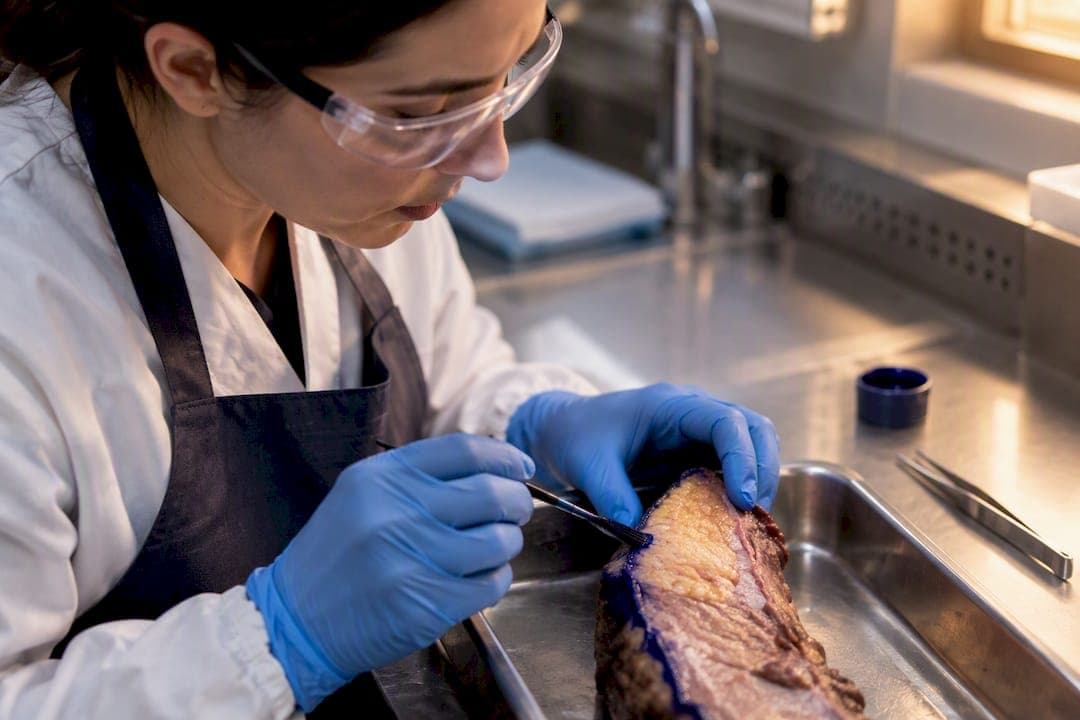
A negative margin means no cancer cells were found at the outer edge of the tissue removed during surgery, while a positive margin means cancer cells extend to that edge. Neither result alone decides your treatment. What happens next depends on the cancer type, how close the tumor came to the edge, and disease-specific guidelines, such as the “no ink on tumor” standard used for invasive breast cancer.
TL;DR:
- Margin status varies significantly by cancer type, with “no ink on tumor” acceptable for breast cancer but strict cutoffs like 1 millimeter for pancreatic cancer.
- Surgical specimen orientation and accurate ink marking are crucial for reliable margin assessment, given the impact on treatment decisions.
- A positive or close margin often leads to additional surgery, radiation, or systemic therapy, but the response depends on tumor biology and overall clinical context.
- Margin results are estimates based on sampled tissue, so they are not foolproof, and ambiguous findings require multidisciplinary review.
- Clear communication and thorough review of pathology reports help prevent delays and ensure appropriate treatment planning.
Table of Contents
- What Does Margin Status Mean in Pathology?
- How Pathologists Assess and Report Margins
- Do Margin Standards Differ by Cancer Type?
- What Happens After a Positive or Close Margin?
- What Does the Evidence Say About Margins and Recurrence?
- How Should I Read My Pathology Report?
- Why Margin Assessment Has Limits
- How Laboratory Practices Shape Margin Reporting
- Get Clarity on Your Pathology Results
- Sources
- FAQ
What Does Margin Status Mean in Pathology?
When a surgeon removes a tumor, the pathologist examines the outer rim of that tissue under a microscope. That rim is the surgical margin, and how close cancer cells sit to its edge determines the margin status pathology teams report back to your surgical team.
The National Cancer Institute defines the margin as the edge of tissue removed during cancer surgery. A pathologist assigns one of three labels after reviewing that edge under the microscope:
- Negative (clean or clear): No cancer cells appear at the cut edge, meaning the surgeon likely removed the entire visible tumor plus a rim of normal tissue.
- Positive (involved): Cancer cells touch the very edge of the removed tissue, meaning some tumor may remain in the patient’s body.
- Close: Cancer cells sit near the edge but not touching it, usually measured in millimeters rather than described as clearly clean.
Pathologists sometimes use “at ink” to describe cells touching the inked surface directly, versus “near ink” for cells sitting a measured distance away. Some reports also use R-classification, borrowed from surgical oncology: R0 means no residual tumor, R1 means microscopic residual disease at the margin, and R2 means visible residual tumor left behind. Not every lab uses R-classification routinely, so don’t be alarmed if your report skips it entirely.
How Pathologists Assess and Report Margins
The number that ends up on your report starts long before anyone looks through a microscope. It starts in the grossing room, where a pathology assistant paints the outer surface of the specimen with colored ink, often using different colors for different sides so the surgical team knows exactly which margin is which.
Orientation matters enormously here. Surgeons frequently place a suture or clip on one edge of the tissue to mark “superior” or “lateral,” and that marker tells the lab which margin corresponds to which direction in the patient’s body. A misplaced or missing suture can turn a routine case into a confusing one, since the pathologist has no reliable way to say which edge came closest to disease.
Once the specimen is inked and sectioned, the pathologist measures the closest distance between tumor cells and the inked surface, typically in millimeters. That distance gets recorded for each named margin, since a tumor near the deep margin behaves differently than one near a lateral or superficial margin.
Here’s roughly how a specimen moves from operating room to final report:
- The surgeon submits the specimen, sometimes with orientation sutures or clips attached.
- The grossing pathologist or assistant inks each margin surface and takes measured sections.
- Additional shave margins, if submitted separately, get their own labels and independent assessment.
- The pathologist reviews slides microscopically and measures the closest tumor-to-ink distance.
- The final report lists margin status, distance, and location for each named margin.
Separately submitted shave margins can complicate the picture. If a surgeon takes an extra shave from the tumor bed after the main specimen comes out, that shave gets its own read, and a positive main-specimen margin paired with a negative shave margin often changes the overall clinical picture.
Pro Tip: Look for four things on your report: the final margin status line, the measured distance in millimeters, which specific margin was involved, and the specimen map showing where each section came from. Those four fields together tell you far more than the summary line alone.
Do Margin Standards Differ by Cancer Type?
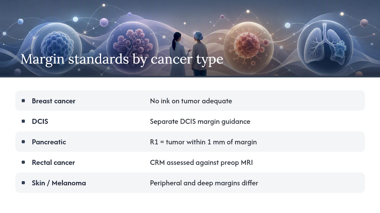
Yes, and this is where a lot of confusion creeps in. A margin distance considered perfectly fine for breast cancer would be alarming for pancreatic cancer, because each tissue type and surgical approach carries its own risk profile.
Breast cancer follows one of the clearer standards in oncology. The SSO–ASTRO consensus guideline states that “no ink on tumor” is an adequate margin for stage I or II invasive breast cancer treated with breast-conserving surgery followed by whole-breast radiation. Wider margins don’t reliably improve local control in that setting, which runs counter to what many patients assume. Ductal carcinoma in situ carries its own separate margin recommendations, since it behaves differently than invasive disease.
Pancreatic cancer plays by different rules entirely. Much of the literature on pancreatic resection margins treats an R1 (microscopically positive) resection as tumor within 1 millimeter of the margin, and R1 resections tend to associate with worse overall and disease-free survival.
Rectal cancer relies heavily on the circumferential resection margin, or CRM, which surgeons and radiologists correlate closely with preoperative MRI findings before ever reaching the operating room.
Melanoma and other skin cancers separate peripheral margins from deep margins, since depth of invasion carries independent prognostic weight beyond the horizontal spread pathologists measure at the skin’s edge.
The core lesson: never apply a breast-margin rule to a pancreatic case, or a skin-cancer depth rule to a rectal specimen. Site-specific thresholds exist because tumor biology, surgical anatomy, and available adjuvant treatments differ enough that one universal number would mislead more than it would help.
What Happens After a Positive or Close Margin?
A positive margin doesn’t automatically send a patient back to the operating room. Clinicians weigh several factors before deciding on next steps, and that decision-making process is exactly why margin status functions as one input among several rather than a standalone verdict.
Typical responses to a positive or close margin include:
- Re-excision surgery, removing additional tissue from the involved margin site.
- Adjusted radiation planning, sometimes adding a boost dose to the tumor bed instead of returning to surgery.
- Systemic therapy adjustments, such as starting or intensifying chemotherapy or targeted treatment.
- Continued observation, appropriate when the involved area is small and other risk factors are favorable.
The decision hinges on tumor biology, disease stage, imaging findings, and whether the surgeon submitted separate shave margins that came back clean. Patient preference matters too. A patient facing significant surgical risk from a second operation might reasonably choose radiation intensification over re-excision, particularly when the positive margin is small and isolated.
This is also why multidisciplinary tumor boards exist. Surgeons, radiation oncologists, medical oncologists, and pathologists often review the same case together precisely because a single margin finding rarely tells the whole story on its own. Not every positive margin represents equal risk, and not every case warrants the same response.
What Does the Evidence Say About Margins and Recurrence?
The numbers behind margin guidelines come from large pooled analyses, not single studies. A study on surgical margin status and oncologic outcomes in breast-conserving surgery found pooled estimates indicating that a notable minority of cases involve tumor directly on ink or within a close margin distance. Residual disease after re-excision shows up in a meaningful share of cases, particularly when the original tumor was multifocal or carried a substantial DCIS component.
| Finding | Reported Figure | Context |
|---|---|---|
| Tumor on ink (pooled estimate) | ~9.4% | Breast-conserving surgery cohorts |
| Tumor within 2 mm (pooled estimate) | 9.4% | Breast-conserving surgery cohorts |
| Pancreatic R1 margin | Tumor within 1 mm | Associated with worse overall and disease-free survival |
Pancreatic surgery tells a starker story. R1 resections consistently associate with shorter overall survival and shorter disease-free survival compared with R0 resections, which is part of why surgeons push hard for wider clearance when the tumor’s location allows it.
None of these figures translate cleanly across eras or institutions. Older studies predate modern radiation techniques and systemic therapies that now compensate for narrower margins, and pooled estimates mix different surgical techniques, tumor grades, and follow-up periods. A 9.4% rate of tumor on ink from a multi-institution meta-analysis reflects an average, not a prediction for any individual patient. Treat these figures as a snapshot of population-level risk, useful for understanding why guidelines exist, rather than a personal forecast.
How Should I Read My Pathology Report?
Reading a pathology report feels intimidating the first time, mostly because the terminology looks nothing like everyday language. A few sections deserve your attention above the rest.
- Find the final diagnosis line first. This usually states margin status in one sentence, often the fastest way to get the headline result.
- Check the measured distance. Look for millimeters listed near the margin description, since “close” can mean anywhere from 0.1 mm to several millimeters depending on the lab.
- Identify which margin was involved. A positive posterior margin carries different implications than a positive superficial margin, depending on tumor location.
- Read the margin comment section. Pathologists frequently add context here explaining ambiguous findings or noting separately submitted shave margins.
- Review the specimen map if one is included. This diagram shows where each tissue section came from, which helps your surgeon interpret the finding spatially.
Bring specific questions to your next appointment: Which margin was involved, and how close was it? Were separate shave margins submitted, and what did they show? Does my case need multidisciplinary review before we decide on treatment?
Pro Tip: If your report reads unclear, contradicts your imaging, or uses unfamiliar terminology, ask your care team about a second pathology opinion or digital review before treatment decisions move forward. A second set of eyes catches more than people expect.
Why Margin Assessment Has Limits
Margin status isn’t a perfect measurement, and understanding its limits keeps patients and clinicians from over-reading a single result. Pathologists examine representative sections of a specimen, not its entire surface, which means a clean margin on the slides reviewed doesn’t guarantee zero cancer cells exist anywhere along that edge.
A few common pitfalls show up repeatedly:
- Fragmented or piecemeal specimens make orientation difficult, since a surgeon removing tissue in pieces loses the clear anatomical reference an intact specimen provides.
- Sampling limitations mean the lab examines a subset of tissue sections, not the entire cut surface, so extremely rare or scattered cells could theoretically go undetected.
- Institutional template differences mean one hospital’s report format may present the same finding differently than another’s, which can confuse patients comparing reports.
- “Cannot be determined” findings signal a need for clinical-pathologic correlation, not an assumption of negative or positive status by default.
When a report includes ambiguous or “cannot be determined” language, that’s a cue to loop in the surgeon and pathologist together rather than proceed on assumption.
How Laboratory Practices Shape Margin Reporting
Margin status pathology only helps patients and clinicians when the underlying report is consistent, detailed, and fast enough to inform treatment timing. That consistency depends heavily on lab practices most patients never see.
Margin reporting can be structured around board-certified pathologists with subspecialty expertise, working from accredited protocols that call for site-specific fields rather than a one-size-fits-all template. That structure matters because a breast case and a skin case genuinely need different reporting frameworks, not the same boilerplate language stretched to fit both.
Digital pathology tools also change how quickly a second reviewer can weigh in on a borderline call, since slides can be reviewed remotely without waiting for physical shipment between facilities. For patients or clinicians facing an ambiguous margin finding, that speed and access to subspecialty review can shape how quickly a treatment plan moves forward.
— EIV Diagnostics
Get Clarity on Your Pathology Results
Margin status pathology reports carry weight, and getting a clear, well-documented read the first time saves patients from delays and second-guessing later. Patients can access pathology services across histopathology, digital pathology, and molecular pathology, with the option for mobile phlebotomy so sample collection can happen at home or in the office.

If your report feels unclear, or your surgical team wants a subspecialty second read before finalizing treatment, EIV Diagnostics offers digital pathology review alongside standard histopathology services. Providers and self-pay patients can also explore the full range of diagnostic services, including molecular pathology testing, on the complete services page. Individuals may request a report review or schedule mobile phlebotomy for follow-up testing via the provider’s website.
Sources
- NCI Dictionary of Cancer Terms — Margin
- SSO–ASTRO consensus guideline on margins for breast-conserving surgery
- Relevant pancreatic margin literature (PubMed listing)
This article is general information, not a substitute for advice from a qualified doctor. Consult a qualified healthcare professional about your own circumstances before acting on anything here.
FAQ
What Does a Positive Margin Mean?
A positive margin means cancer cells were found at the very edge of the tissue the surgeon removed, suggesting some tumor may remain in the body. It doesn’t automatically mean another surgery is required. The decision depends on which margin was involved, how much tissue was affected, and disease-specific factors your care team reviews together.
What Does “Negative for Invasive Carcinoma” Mean on a Margin?
This phrase means the pathologist found no invasive cancer cells at that specific margin’s edge, though it says nothing about other margins on the same specimen. Always check whether the report addresses each named margin separately, since one clean margin doesn’t guarantee all margins are clean.
Do You Need Radiation If Margins Are Clear?
A clear margin doesn’t automatically remove the need for radiation. For invasive breast cancer treated with breast-conserving surgery, the SSO–ASTRO guideline still generally recommends whole-breast radiation even after a negative “no ink on tumor” margin, since radiation and margin clearance serve different roles in reducing recurrence risk.
What Does It Mean When a Surgical Margin Is Positive?
It means the pathologist identified tumor cells at the outer inked edge of the removed specimen during microscopic examination. Clinicians typically respond with re-excision, adjusted radiation planning, or systemic therapy, chosen based on tumor type, location, and multidisciplinary review rather than a fixed rule.