
By EIV Diagnostics · July 7, 2026
Dermatopathology Services: What Patients and Providers Need to Know
Discover how dermatopathology services provide accurate skin disease diagnoses. Learn the benefits for patients and providers in Fresno.
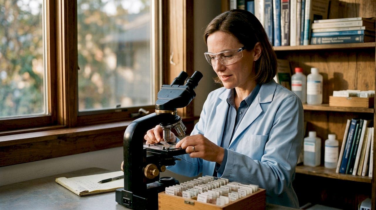
Dermatopathology services are defined as the specialized microscopic examination of skin tissue samples to diagnose skin diseases with clinical precision. A board-certified dermatopathologist analyzes biopsied tissue at the cellular level, correlating microscopic findings with clinical data to reach a definitive diagnosis. These services cover more than 1,500 recognized skin conditions, from inflammatory disorders to melanoma. For patients and providers in Fresno, understanding how this process works means faster, more confident treatment decisions.
What dermatopathology services actually involve
Dermatopathology is the subspecialty of medicine that sits at the intersection of dermatology and pathology. The formal industry term is dermatopathology, and it refers specifically to laboratory-based analysis of skin tissue rather than clinical skin exams performed in an office. A dermatopathologist does not typically see patients directly. Instead, they receive tissue samples collected by dermatologists or surgeons, examine them under a microscope, and produce a written diagnostic report.
The scope of skin pathology services is broader than most patients realize. A dermatopathology laboratory handles everything from routine mole checks to complex autoimmune blistering diseases and rare cutaneous lymphomas. The diagnostic report produced by the lab directly shapes whether a patient receives surgery, topical therapy, systemic medication, or watchful monitoring. Getting that report right is not optional. It is the foundation of every treatment decision that follows.
Dermatopathologists correlate clinical suspicion from the referring dermatologist with microscopic tissue findings to produce reliable diagnoses. This integrative process is what separates dermatopathology from a simple lab test. The pathologist functions as a diagnostic partner, not just a technician reading slides.
What training and expertise do dermatopathologists have?
Becoming a board-certified dermatopathologist requires 13–15 years of training after college, including medical school, a residency in either dermatology or pathology, and a dedicated dermatopathology fellowship. That training timeline is longer than most surgical specialties. The depth reflects how technically demanding the work is.
There are two main training paths into dermatopathology:
- Dermatology residency route: The physician completes a dermatology residency, then pursues a one-year dermatopathology fellowship. This path produces specialists with strong clinical skin disease knowledge.
- Pathology residency route: The physician completes anatomic pathology training, then completes the same fellowship. This path produces specialists with deep expertise in tissue processing, staining, and laboratory science.
Both routes lead to the same board certification examination administered by the American Board of Dermatology and the American Board of Pathology. Either credential qualifies the physician to practice dermatopathology at the same standard.
The core technical skills required include advanced microscopy, pattern recognition across hundreds of disease presentations, and proficiency in ancillary testing methods. Molecular and immunohistochemical tests are selectively applied based on clinical relevance and microscopic ambiguities to refine a diagnosis. A dermatopathologist must know when a standard hematoxylin and eosin stain is sufficient and when additional testing is necessary.
Pro Tip: When you send a biopsy to a dermatopathology lab, always include a brief clinical note describing the lesion’s appearance, location, and duration. This context directly improves diagnostic accuracy because the pathologist correlates your clinical impression with what they see under the microscope.
How is a skin biopsy processed and analyzed in a dermatopathology lab?
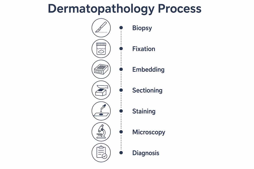
The journey from biopsy to diagnosis follows a precise, multi-step workflow. Each step affects the quality of the final result.
-
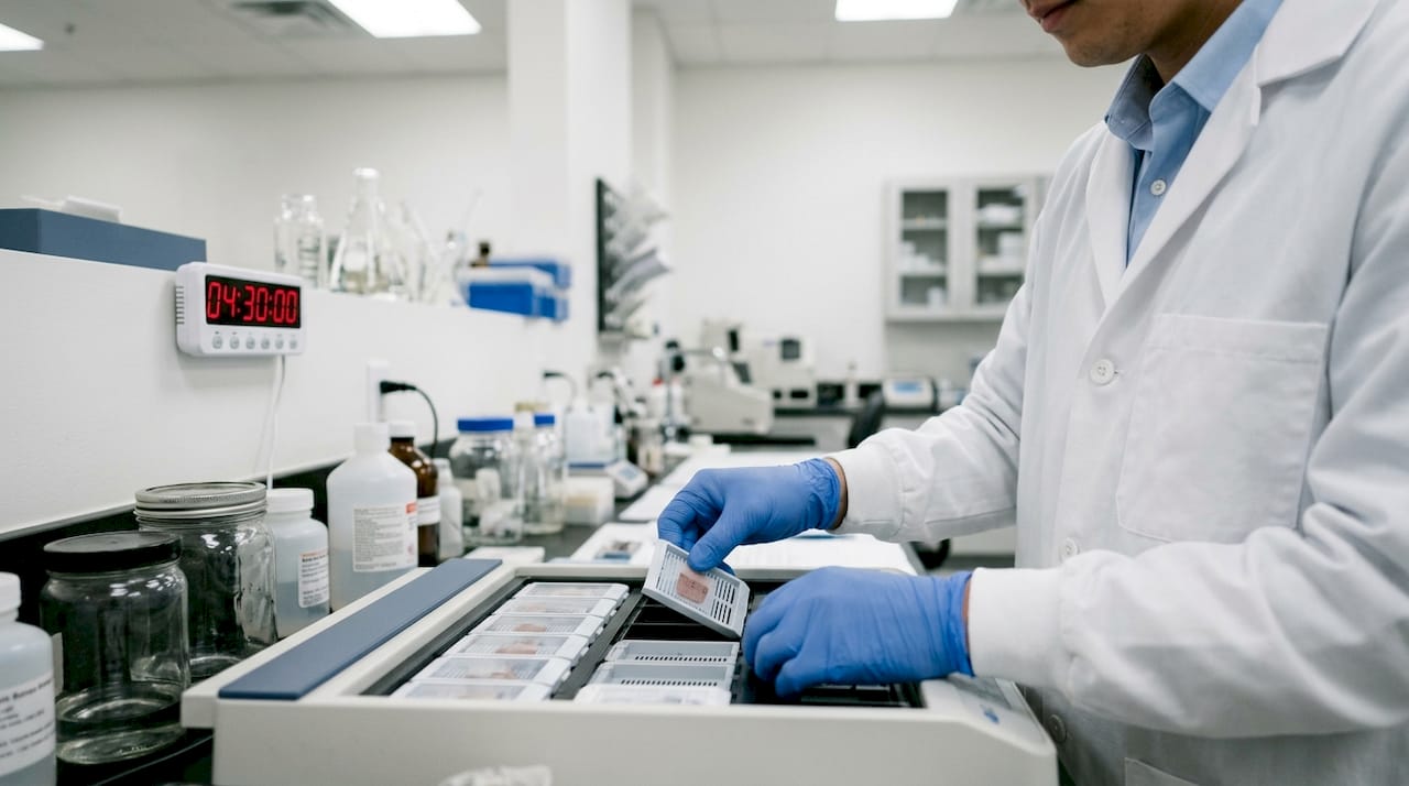
Fixation. The tissue sample is placed in formalin immediately after collection. Proper fixation in formalin is critical. Delays or inadequate fixation cause autolysis, a process where cellular structures break down and become unreadable. A compromised sample can prevent a definitive diagnosis entirely.
-
Processing. The fixed tissue is dehydrated through a series of alcohol solutions, then cleared with a solvent, and finally infiltrated with liquid paraffin wax. This process makes the tissue firm enough to cut into extremely thin sections.
-
Embedding and orientation. The tissue is embedded in a paraffin block. The orientation of the specimen during embedding determines which tissue planes the pathologist can examine. Incorrect orientation can hide critical diagnostic features. This step requires skilled histotechnologists who understand tissue anatomy.
-
Microtomy. A microtome slices the paraffin block into sections at approximately 4 micrometers thickness, a fraction of the width of a human hair. These ultra-thin sections are mounted onto glass slides.
-
Staining. Hematoxylin and eosin staining is the standard method for highlighting cellular structures on biopsy slides. H&E staining colors cell nuclei blue and cytoplasm pink, making tissue architecture visible. For complex or ambiguous cases, immunohistochemistry adds protein-specific markers that identify cell types or tumor markers.
-
Microscopic interpretation. The dermatopathologist examines the stained slides, integrates the clinical history, and renders a diagnosis. This is the step where pattern recognition and clinical correlation determine the final report.
| Processing step | Purpose | Key risk if skipped or rushed |
|---|---|---|
| Formalin fixation | Preserves tissue architecture | Autolysis destroys cellular detail |
| Paraffin embedding | Creates cuttable tissue block | Poor orientation hides diagnostic areas |
| Microtomy at 4 micrometers | Produces readable thin sections | Thick sections obscure cell morphology |
| H&E staining | Highlights cellular structures | Tissue remains invisible under microscope |
| Immunohistochemistry | Identifies specific cell markers | Ambiguous diagnoses remain unresolved |
Pro Tip: If you are a provider submitting biopsies for dermatological testing, confirm that your collection protocol specifies immediate formalin fixation. A 30-minute delay in fixation at room temperature can degrade tissue quality enough to affect the diagnostic report.
What clinical value and turnaround times can patients and providers expect?
A dermatopathology report does far more than name a diagnosis. Detailed reports inform treatment, staging, and follow-up decisions by including tumor thickness, margin status, and notes on whether additional testing is recommended. For melanoma, the Breslow thickness measurement in the report directly determines the surgical margin required and whether sentinel lymph node biopsy is indicated. The report is a clinical roadmap, not just a label.
Routine results from a dermatopathology lab are typically available within 3–5 business days. Urgent findings, such as a suspected melanoma or high-grade malignancy, are communicated directly by phone to the ordering provider. That distinction matters because it means providers do not need to check a portal repeatedly for critical results.
Providers can expect results delivered through several channels:
- Electronic health record integration: Reports are uploaded directly into the provider’s EHR system, reducing manual data entry and transcription errors.
- Fax transmission: Still widely used for providers without full EHR connectivity, particularly in smaller Fresno-area practices.
- Direct phone calls: Reserved for urgent or unexpected findings that require immediate clinical action.
- Written pathology reports: Formal documents that include all diagnostic details, ancillary test results, and pathologist commentary.
One common misconception is that patients need to obtain physical glass slides to get a second opinion or consultation. The finalized pathology report contains sufficient information for an educational consultation. Physical slides are only necessary when a formal diagnostic re-interpretation is requested.
How do dermatopathology consultations and second opinions work?
Second opinions in pathology for skin diseases fall into two distinct categories, and confusing them leads to delays and frustration.
A diagnostic second opinion is a formal re-analysis of the original glass slides by a different board-certified dermatopathologist. This involves slide re-interpretation and produces a new independent diagnostic report. Providers typically request this when the original diagnosis is ambiguous, when the clinical picture does not match the pathology report, or when a rare diagnosis requires subspecialty confirmation.
An educational consultation is different. It focuses on explaining and clarifying an already finalized pathology report without reviewing the original slides. Patients do not need to submit physical slides for this type of consultation. The written report alone provides the information needed to answer questions about what the diagnosis means, what the terminology refers to, and what treatment implications follow.
Knowing which type of consultation you need saves time and reduces the burden on patients who may already be managing a stressful diagnosis. For providers, it also clarifies what to request from the laboratory and what documentation to prepare.
When additional molecular or immunohistochemical testing is warranted, the dermatopathologist will note this in the original report. Providers should act on those recommendations promptly, as ancillary testing results can change the diagnosis or refine the staging. Eivdiagnostics offers molecular pathology services that support exactly this kind of follow-on testing for complex skin disease cases.
Key Takeaways
Accurate dermatopathology diagnosis depends on proper biopsy handling, board-certified expertise, and clear communication between the lab and the ordering provider.
| Point | Details |
|---|---|
| Training is extensive | Board-certified dermatopathologists complete 13–15 years of training before independent practice. |
| Fixation is critical | Immediate formalin fixation after biopsy collection is required to preserve tissue for accurate analysis. |
| Reports guide treatment | Dermatopathology reports include tumor thickness, margin status, and ancillary test notes that directly shape clinical decisions. |
| Turnaround is predictable | Routine results arrive within 3–5 business days; urgent findings are communicated by phone. |
| Second opinions have two forms | Diagnostic re-interpretation requires slides; educational consultations require only the finalized report. |
Why dermatopathology deserves more attention than it gets
Working closely with pathology cases over the years, I have noticed that dermatopathology is consistently underestimated by both patients and referring providers. Most people think of a biopsy as a simple procedure with a straightforward result. The reality is that the laboratory process is where the real diagnostic work happens, and small errors at any stage can cascade into a wrong or delayed diagnosis.
The most underappreciated factor is biopsy orientation during embedding. I have seen cases where a technically well-collected specimen produced an uninformative report simply because the tissue was embedded at the wrong angle. The pathologist could only see a cross-section of tissue that missed the diagnostic area entirely. This is not a failure of the pathologist. It is a failure of the pre-analytical process that most clinicians never think about.
Digital pathology is changing this. Whole-slide imaging allows pathologists to review cases remotely, share images with colleagues instantly, and apply computational tools to flag areas of interest. Eivdiagnostics has invested in digital pathology capabilities that make this kind of collaboration practical for Fresno-area providers who need specialist input quickly. The technology does not replace the pathologist’s judgment. It gives that judgment better tools and faster access to expertise.
The best outcomes I have observed come from practices where the dermatologist and dermatopathologist communicate directly, not just through reports. A brief call to discuss a borderline case often resolves ambiguity faster than ordering additional tests. That relationship is worth building.
— Krunal
Eivdiagnostics dermatopathology services for Fresno providers and patients
Eivdiagnostics is an independent pathology laboratory in Fresno, CA, built to serve both healthcare providers and patients who need reliable skin pathology services without the delays of large institutional labs.

The lab’s dermatopathology services include H&E analysis, immunohistochemistry, and molecular testing for complex skin disease cases. Board-certified pathologists produce detailed reports with fast turnaround times, delivered through EHR integration or direct provider communication. Patients with a prescription and self-pay patients are both welcome. For providers managing complex dermatology cases in the Central Valley, Eivdiagnostics offers a direct line to expert diagnostic support. Contact Eivdiagnostics to discuss your diagnostic needs or submit a case for review.
FAQ
What is dermatopathology?
Dermatopathology is the medical subspecialty that diagnoses skin diseases by examining biopsied tissue under a microscope. A board-certified dermatopathologist correlates microscopic findings with clinical data to produce a diagnostic report.
How long does a dermatopathology report take?
Routine dermatopathology results are available within 3–5 business days. Urgent findings are communicated directly by phone to the ordering provider.
Do I need physical slides for a second opinion?
Physical slides are required only for a formal diagnostic re-interpretation. An educational consultation that explains your existing report requires only the finalized pathology document.
What stains do dermatopathology labs use?
Hematoxylin and eosin staining is the standard method for all skin biopsies. Immunohistochemical stains are added selectively when the H&E findings are ambiguous or when specific cell markers need confirmation.
Can patients in Fresno access dermatopathology services directly?
Yes. Eivdiagnostics in Fresno accepts patients with a provider prescription and self-pay patients who want direct access to pathology testing without a referral requirement.