
By EIV Diagnostics · October 3, 2026
6–72 Hour Fixation Window: Evidence First Formalin Protocols for Labs
Evidence-first lab reference on formalin fixation: keep cold ischemia under 60 minutes, use a 6–72 hour fixation window, and follow OSHA safety and...
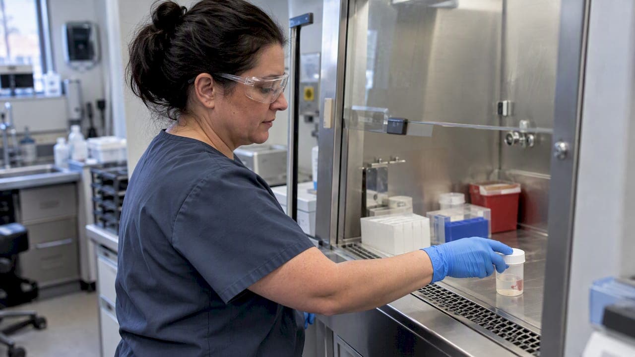
Keep cold ischemia time under 60 minutes before fixation begins, and handle formalin under OSHA formaldehyde controls at every step.
TL;DR:
- Maintaining cold ischemia time under 60 minutes before fixation is crucial to preserve biomarker integrity and ensure accurate testing results.
- Using 10% neutral buffered formalin with a 10:1 formalin-to-tissue ratio and proper specimen slicing improves fixation quality and histological accuracy.
- Proper documentation of fixation start time and cold ischemia duration supports reliability and defensibility of downstream diagnostic results.
- Validating alternative fixatives requires parallel testing, revalidation of assays, and thorough documentation before replacing formalin in routine practice.
- OSHA compliance, including safety controls and exposure monitoring, remains essential for laboratories working with formalin to protect staff and meet regulatory standards.
Table of Contents
- Why 10% neutral buffered formalin is the standard fixative
- Setting up and handling formalin in the biopsy workflow
- How fixation timing affects IHC and molecular test results
- Preparing 10% neutral buffered formalin correctly
- Workplace safety and OSHA formaldehyde compliance
- When to consider alternatives to formalin fixation
- How EIV Diagnostics approaches fixation and specimen quality
- Where EIV Diagnostics fits into your biopsy workflow
- Balancing archival habit with molecular-era demands
- Sources
- FAQ
Why 10% neutral buffered formalin is the standard fixative
Formalin is a water-based solution of formaldehyde gas, and 10% neutral buffered formalin is that solution diluted to roughly 4% formaldehyde by weight, buffered to a neutral pH with phosphate salts. This combination cross-links proteins fast enough to stop tissue autolysis while leaving nuclear and cytoplasmic detail readable under routine staining. The phosphate buffer matters more than lab staff often assume: an acidic or unbuffered formalin solution can form formalin pigment deposits and distort nuclear morphology, which complicates both diagnosis and downstream antigen detection.
Most commercial 10% NBF includes a small amount of methanol as a stabilizer to slow paraformaldehyde formation during storage. That additive has little effect on routine histology but is worth knowing about if a lab validates a fixative against a strict chemical specification, since some molecular assays specify methanol-free formalin.
The continued dominance of 10% NBF is less about proving it outperforms every alternative and more about decades of accumulated clinical and archival data. A review of formalin fixation and tissue processing describes 10% NBF as the universal routine fixative for paraffin-embedded sections, a status built on established practice rather than head-to-head superiority over every niche fixative. That matters for any lab considering a change: switching away from NBF means losing direct comparability with decades of archived slides and published reference ranges.
Key properties that make 10% NBF the default choice:
- Penetration and cross-linking speed that works across most routine specimen sizes without specialized equipment.
- Neutral pH buffering that limits formalin pigment and preserves nuclear detail.
- Compatibility with routine H&E staining, most immunohistochemistry panels, and many molecular assays after appropriate fixation time.
- Archival continuity, since most reference literature and retrospective studies were generated on NBF-fixed tissue.
Setting up and handling formalin in the biopsy workflow
Getting the ratio and timing right starts the moment a specimen leaves the patient. A 10:1 formalin to tissue volume ratio gives the fixative enough volume to maintain concentration as it diffuses into the tissue instead of being depleted near the specimen surface. For a biopsy that displaces roughly 2 milliliters of fixative, that means about 20 milliliters of formalin in the container, and larger excisions need proportionally more.
Thin or dense specimens fix unevenly if left whole. Core needle biopsies generally fix well as submitted, but larger excisions benefit from sectioning into slices around 4 to 5 millimeters thick so formalin can reach the center before autolysis sets in.
A practical sequence for receiving and fixing a specimen:
- Confirm the specimen container holds the correct fixative and that volume meets the 10:1 ratio before adding tissue.
- Inspect prefilled formalin containers for cracks, loose caps, or visible leakage before use, since adverse event reports have documented leaking prefilled formalin containers reaching labs from at least one major supplier.
- Section larger specimens to expose adequate surface area without compromising margins or orientation.
- Record the time the specimen left the patient and the time it entered formalin, since that interval is the cold ischemia time.
- Label the container with patient identifiers, specimen source, and date and time of fixation start, then log it against the requisition.
Pro Tip: Keep a simple time-stamped log at the grossing station so cold ischemia time and fixation start time are recorded automatically rather than reconstructed later.
Documentation gaps are one of the most common and most avoidable sources of downstream dispute, particularly when a biomarker result is questioned months after the original diagnosis. A written fixation record turns a guess into a defensible fact. For broader processing workflow reminders, see this guide to biopsy processing steps.
How fixation timing affects IHC and molecular test results
Cold ischemia time, the interval between tissue devascularization and the start of fixation, has a direct bearing on biomarker reliability. ASCO and CAP joint preanalytic recommendations call for cold ischemia time under 60 minutes and a formalin fixation time generally between 6 and 72 hours, a window intended to optimize immunohistochemistry reliability for markers used in breast cancer workup.

A timed fixation study found that estrogen receptor IHC staining scores plateau at 6 to 8 hours of formalin exposure, which is the basis for the commonly cited minimum fixation time for reliable ER results. Below that threshold, cross-linking is incomplete and staining becomes inconsistent across the specimen; above the 72 hour mark, prolonged fixation risks masking epitopes and over-cross-linking nucleic acids, which can reduce antibody binding and complicate molecular testing that relies on amplifiable DNA or RNA.
Under-fixation and over-fixation cause different but equally disruptive problems:
- Under-fixed tissue shows patchy or weak staining because formaldehyde has not fully penetrated and cross-linked the center of the specimen.
- Over-fixed tissue can show reduced antigenicity on some epitopes, since extended cross-linking changes protein conformation.
- Both extremes complicate molecular assays that depend on intact, amplifiable nucleic acids, since fixation chemistry affects DNA and RNA quality alongside protein structure.
Not every deviation from the target window changes the result. A retrospective review of endometrial cancer specimens with a median cold ischemia time of 33 minutes found no significant differences in ER, PR, HER2, or mismatch repair protein expression tied to cold ischemia or fixation time variation in that cohort, though the authors noted that ASCO/CAP breast cancer preanalytic standards remain the default reference point pending organ-specific guidance. That finding is a useful caution against treating every minor timing deviation as disqualifying, but it is not a license to ignore the target windows, since breast IHC remains the best-studied case and the one with the clearest timing thresholds.
Fixation quality is now something labs can assess directly rather than infer. Research into real-time fixation quality assessment using ultrasound time-of-flight analysis illustrates how central fixation has become as a preanalytic checkpoint for diagnostic accuracy, even though such tools remain largely investigational rather than routine.
When a specimen falls outside the target window, document the actual cold ischemia time and fixation duration on the report rather than omitting the detail, and flag any biomarker result generated from a specimen with known preanalytic deviation so the ordering clinician can weigh it appropriately. More detail on how fixation duration affects staining appears in this explainer on immunohistochemistry and fixation timing.
Preparing 10% neutral buffered formalin correctly
Diluting it to 10% NBF means mixing one part of that stock with nine parts of phosphate-buffered water, which yields a working solution containing roughly 3.7% to 4% actual formaldehyde, the concentration referred to as 10% NBF in histology practice.
A practical preparation sequence:
- Start with 37 to 40% formaldehyde stock and dilute it 1 part stock to 9 parts buffered water to produce working 10% NBF.
- Use a monobasic and dibasic sodium phosphate buffer combination to hold the solution near neutral pH, then verify with a pH meter or strip before use in a new batch.
- Store prepared formalin at room temperature in a sealed, labeled container away from direct sunlight, and discard solutions showing cloudiness or precipitate.
- Check expiration dating on commercial prebuffered formalin and rotate stock so older lots are used first.
That tradeoff, slightly higher cost for better traceability, is usually worth it for any lab operating under accreditation review.
Workplace safety and OSHA formaldehyde compliance
The standard requires employers to implement exposure controls, employee training, hazard labeling, and personal protective equipment provisions wherever formaldehyde-containing materials are used.
OSHA’s standard also requires medical surveillance under specific exposure conditions, which means labs with higher formalin throughput need a documented monitoring plan, not just protective equipment at the bench.
Baseline controls every lab handling formalin should have in place:
- Nitrile gloves, splash goggles, and a fitted respirator or local exhaust ventilation when working with open containers of formalin.
- Safety data sheets accessible at the point of use, not just filed in an office binder.
- Written spill response procedures with absorbent material and neutralizing agents staged near grossing and processing stations.
- Labeled waste containers for formalin-contaminated material, disposed of through a permitted hazardous waste handler rather than down the drain.
Exposure monitoring becomes necessary when air sampling or job task review suggests employees may approach or exceed OSHA’s action level, and any lab that has not run a baseline exposure assessment in a formalin-heavy grossing area should treat that as overdue rather than optional. Accreditation inspections frequently check these exact controls, and a gap here is one of the more common citations labs receive. For a broader look at readiness, see this guide to pathology lab accreditation and inspection readiness.
When to consider alternatives to formalin fixation
Formalin is not the only fixative in use, and some specimen types are better served by something else. Alcohol-based fixatives preserve nucleic acids better for certain molecular panels but can distort cytoplasmic detail. Zinc-based fixatives avoid formaldehyde cross-linking artifacts and work well for some immunofluorescence applications. Cryopreservation, freezing tissue instead of chemically fixing it, is the preferred approach for frozen section diagnosis and for assays that require unfixed, intact nucleic acids or proteins. Glutaraldehyde-based fixatives remain standard for electron microscopy because they preserve ultrastructural detail formalin cannot match.
None of these alternatives carries formalin’s depth of archival and comparative data, which is the real reason formalin remains dominant rather than any single superior property.
Before adopting an alternative fixative for routine use, a lab should work through a validation checklist:
- Run parallel fixation on split specimens using both the current and proposed fixative to compare morphology and staining.
- Revalidate each antibody or molecular assay against the new fixative rather than assuming existing validation transfers.
- Include known positive and negative controls in every validation run to confirm the new fixative does not introduce false results.
- Document the validation study for accreditation review before the new fixative enters routine use.
How EIV Diagnostics approaches fixation and specimen quality
An independent pathology laboratory offering histopathology, immunohistochemistry, molecular pathology, and digital pathology services, staffed by board-certified pathologists who review fixation quality as part of routine case workup.
Operational practices that support reliable results:
- Container inspection on receipt, checking for leaks or damage before a specimen is logged in, consistent with the prefilled-container cautions documented in device adverse event reports.
- Time-stamped documentation of cold ischemia time and fixation duration tied to each requisition, so any biomarker result can be traced back to its preanalytic handling.
- Assay-specific validation before a test is offered clinically, rather than assuming a protocol validated on one specimen type transfers automatically to another.
Providers and labs evaluating a submission partner for formalin-fixed specimens can review EIV Diagnostics’ histopathology services for details on processing and turnaround.
Where EIV Diagnostics fits into your biopsy workflow

Getting fixation right is only half the preanalytic picture. Specimen transport and cold chain handling before formalin even enters the picture matter just as much, and this specimen cold chain management guide covers that side well.
- Histopathology, immunohistochemistry, and molecular pathology run under board-certified pathologist oversight with documented fixation and validation records.
- Mobile phlebotomy brings specimen collection to a patient’s home or office, reducing the handling gaps that can extend cold ischemia time.
- Digital pathology supports remote case review and consultation without delaying turnaround.
If your lab or practice needs a validated partner for formalin-fixed specimen processing, visit EIV Diagnostics’ services to start a submission or request a consultation.
Balancing archival habit with molecular-era demands
Formalin earned its place through decades of consistency, not because it is the only fixative that works. The honest tension in any lab today is between that archival continuity and the sharper preanalytic demands of molecular assays, which punish sloppy timing in ways routine H&E never did. Before changing a fixation protocol, validate it in parallel, document the comparison, and loop in both the clinical team ordering the test and the lab running it. Coordination beats assumption every time a result sits close to a threshold.
— EIV Diagnostics
This article is general information, not a substitute for advice from a qualified doctor. Consult a qualified healthcare professional about your own circumstances before acting on anything here.
Sources
The guidance in this article draws on a small set of primary sources worth keeping on hand for protocol review or accreditation documentation.
- Formalin fixation and tissue processing for diagnostic pathology (review)
- Minimum formalin fixation time for consistent estrogen receptor immunohistochemical staining of invasive breast carcinoma
- 1910.1048 - Formaldehyde. | Occupational Safety and Health Administration
FAQ
How long can a biopsy stay in formalin?
A biopsy can generally stay in 10% neutral buffered formalin for up to 72 hours without significant loss of staining quality, with a practical minimum of 6 to 8 hours for reliable immunohistochemistry results. Specimens left well beyond that window risk epitope masking and reduced antibody binding on certain markers.
What are the five fixatives used in histopathology?
Each serves a different combination of morphology preservation and downstream assay compatibility.
What is the purpose of formalin in biopsy procedures?
Formalin stops tissue autolysis and cross-links cellular proteins so the specimen’s structure stays intact for sectioning, staining, and diagnostic review. This fixation step is what allows a biopsy taken from a patient to be processed into a stable slide days or weeks later.
How do I prepare 10% formalin for histology?
Many labs instead purchase commercially prebuffered 10% NBF to reduce preparation error and get documented concentration and pH on each lot.